


REVIEWS | SMALL RUMINANTS DISEASES
Introduction. Caprine arthritis-encephalitis (CAE) is a chronic viral disease of goats caused by a lentivirus from the Retroviridae family (small ruminant lentivirus group, SRLV). Infection typically involves prolonged asymptomatic carriage, progressing to debilitating lesions in the joints, central nervous system, lungs, and mammary gland. Transmission occurs primarily via colostrum, milk, and direct contact, including fomites and co-mingling with infected animals. Diagnosis relies on serological and molecular methods.
Objective. To summarize international methodological approaches for the prevention of CAE.
Materials and methods. This review was conducted based on an analysis of publications in peer-reviewed scientific journals, official international guidelines, and reports from key organizations such as the Food and Agriculture Organization and the World Organisation for Animal Health. Literature was identified and selected through searches in major scientific databases, including Web of Science, Scopus, PubMed, Google Scholar, Science Hub, CyberLeninka and others.
Results. Analysis of the literature indicates that the most effective outcomes in controlling CAE have been achieved in countries with mandatory national eradication programs, which integrate comprehensive preventive, diagnostic, and administrative measures (e.g., Norway, Switzerland). In nations with voluntary participation programs (e.g., Australia, New Zealand, Canada, Italy, France), positive results are also evident, particularly when supported by incentive mechanisms and sustained educational outreach. Conversely, in countries lacking coordinated national programs (e.g., Turkey, Brazil, Iran, and most African nations), disease control remains inadequate, and seroprevalence rates are consistently high.
Conclusion. The most effective outcomes in controlling CAE have been achieved in countries with mandatory national eradication programs, which integrate comprehensive preventive, diagnostic, and administrative measures (e.g., Norway, Switzerland).
REVIEWS | AVIAN DISEASES
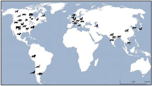
Introduction. In 2024–2025, a number of high pathogenicity avian influenza A(H5N1) outbreaks were reported in dairy cattle. Such an expansion of the range of the virus hosts increases global risks for livestock farming and public health, which requires strengthening animal disease monitoring and control systems.
Objective. Analysis of the epizootological characteristics of avian influenza A(H5N1) virus infection in dairy cattle and other mammals in 2024–2025, as well as summary of the response measures taken and recommendations of international organizations.
Materials and methods. Analytical research methods were used utilizing PubMed, Scopus, Web of Science, Springer, Wiley Online Library databases and materials from international organizations (FAO, EFSA, WOAH, OFFLU, CDC).
Results. Outbreaks of avian influenza A(H5N1) caused by clade 2.3.4.4b virus of genotypes B3.13 and D1.1 in dairy cattle in 2024–2025 occurred as a result of three confirmed independent cases of the pathogen transmission from wild birds. The infection was detected on 1,078 cattle farms in 17 US states. Subsequent virus transmission to poultry, wild and domestic animals, including cats, as well as to humans (70 people), mainly those working on animal and poultry farms, was reported. Reverse transmission of the pathogen from cows back to birds was documented as well. Rapid and wide spread of the virus is associated with extensive animal movements and insufficient biosafety measures. Influenza A(H5) is recommended for the inclusion in the differential diagnosis for cattle, pigs, domestic and wild animals.
Conclusion. Avian influenza A(H5N1) epizootic in cattle and the infection transmission to other mammals pose a serious threat to livestock production and public health. In response to the existing risks, it is necessary to strengthen biosafety measures and surveillance in epidemiologically significant animal populations, incorporate the experience of other countries and establish international cooperation to study the trends of the virus evolution.
REVIEWS | EPIZOOTOLOGY
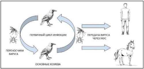
Introduction. West Nile fever is a zoonotic transmissible disease caused by flavivirus that primarily circulates in nature within an enzootic cycle between mosquitoes and birds, and causes disease cases in humans, horses, and other mammals. The rapid expansion of the West Nile fever pathogen range, development of outbreaks with severe clinical manifestations, and the lack of specific preventive tools have been the main arguments for classifying it as a potentially dangerous threat to global health. The close interconnection between human health, animal health, and ecosystems necessitates communication and coordination across the relevant sectors. One Health is an integrated, unifying approach aimed at optimizing the health of humans, animals, and ecosystems involving public health, veterinary and environment protection authorities.
Objective. Analysis of basic epizootological data on the spread of West Nile fever in the Russian Federation.
Materials and methods. The following international and Russian databases were used for literature searching: PubMed, Springer, Google Scholar, CrossRef and Russian Science Citation Index (RSCI), еLibrary, CyberLeninka, respectively. The searching was performed based on the following key words: West Nile fever, One Health, migration of birds, invertebrate hosts, control measures.
Results. In Russia, West Nile virus was first isolated in Astrakhan Oblast in 1963. Currently, presence of the pathogen has been proven in the southern and central regions of the European part of the country, in the south of Western Siberia and the Far East. The lack of disease monitoring in some Russian regions and small numbers of samples tested in most subjects of the Russian Federation hinder an objective assessment of the disease situation, so there is a need to increase the number of tests. It has been shown that the main carriers of the pathogen in our country are mosquitoes of the genera Culex, Anopheles and Aedes, ixodid, argasid and gamasid ticks are also involved in maintaining the virus circulation. The review describes the role of birds in the pathogen transmission, provides data on susceptibility of animals to the infection, discusses modern aspects of West Nile fever diagnosis, prevention and control.
Conclusion. Surveillance of West Nile fever presents a considerable challenge, as the virus circulates among humans, arthropods, and birds. While vaccination is an effective preventive tool, no vaccines against the disease have yet been developed in Russia. In this context, strengthening inter-authority coordination and implementing environmental control measures to limit the virus spread are essential priorities.
ORIGINAL ARTICLES | VETERINARY MICROBIOLOGY
Introduction. Respiratory infections pose a significant challenge in veterinary practice due to their high prevalence across various animal species. Pneumonia and gastrointestinal diseases are leading causes of mortality in captive primates.
Objective. Study of pneumonia incidence in monkeys, the analysis of pulmonary microbiota composition, study of pathomorphological lesions in lung tissue.
Materials and methods. Common methods were used for pathomorphological, microscopic and bacteriological examinations of 1,862 dead monkeys. Lung samples and serum samples from 126 monkeys died of pneumonia in 2021–2024 were tested for acute respiratory viral pathogens as well as for antibodies to them with polymerase chain reaction (PCR) and enzyme-linked immunosorbent assay (ELISA).
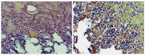
Results. Pneumonia was postmortem diagnosed in 865 monkeys (46.5%). The mortality rate for pneumonia in baby monkeys during their first month of life reached 100%. In baby monkeys under one year of age, the mortality rate was 65.4%. The obtained data showed that the disease incidence in these age groups was high. Deaths of monkeys due to pneumonia were reported throughout the year. Based on postmortem examinations, bilateral polysegmental bronchopneumonia was the most frequent finding, lobar fibrinous pneumonia affecting the right lung was less common. Microscopic analysis detected purulent exudate and cocci bacteria in the bronchial lumen. The predominant bacteria isolated from lung tissue were enterobacteria (58.5%) and Gram-positive cocci (36.6%). Various microorganisms were isolated but the most frequently enteric bacteria were as follows: Escherichia coli (66.1%), Enterococcus spp. (27.5%) and Proteus spp. (31.5%). The following bacterial pathogens associated with pneumonia were detected: Staphylococcus aureus (31.5%), Klebsiella pneumoniae (2.2%), Pseudomonas aeruginosa (0.8%) and Streptococcus pneumoniae (0.6%). Adenoviruses, human parainfluenza viruses of type 1 and type 3 and respiratory syncytial virus (RSV) were also circulated in the monkey colony.
Conclusion. During analysis of microbial etiology of pneumonia in monkeys it shall be considered that pneumonia is frequently arisen as a secondary infection, heavily influenced by underlying gastrointestinal pathologies and immunosuppression.
Introduction. Listeria monocytogenes is a pathogenic microorganism that causes a large number of deaths due to the consumption of contaminated animal products. This underpins the relevance of monitoring the spread of the listeriosis agent in raw materials, animal products, and environmental objects, as well as the antibiotic resistance of isolates.
Objective. Serotype identification and antibiotic resistance analysis of Listeria monocytogenes isolates recovered from animal products in 2021–2024.
Materials and methods. The work was performed at the Microbiology Unit of the Vladimir Testing Laboratory, the Federal Centre for Animal Health. Listeria genus bacteria isolates were identified using time-of-flight mass spectrometry. Antibiotic resistance of isolates belonging to the species Listeria monocytogenes was determined by the disk diffusion method. The values of the growth retardation zones were interpreted according to the Russian recommendations “Determination of the sensitivity of microorganisms to antimicrobial drugs” (IACMAC, version 2025-01). Listeria serological groups were identified using real-time polymerase chain reaction (qPCR) with primers manufactured by Syntol (Russia).
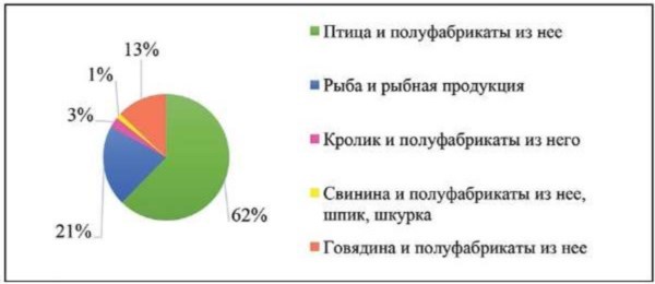
Results. The article presents the results of antibiotic resistance testing of 77 Listeria monocytogenes isolates detected in animal products in 2021–2024, as well as their differentiation by serogroups. Listeria monocytogenes was most frequently detected in poultry products. The detected isolates showed maximum resistance to cefuroxime, sulfamethoxazole/trimethoprim, norfloxacin, rifampicin, levofloxacin, and kanamycin. Moreover, most isolates exhibited resistance to more than one antimicrobial medicinal product. The study established the belonging of the Listeria monocytogenes isolates to the following serogroups: IIa (serotypes 1/2a, 3a) – 92.2%; IIc (serotypes 1/2c, 3c) – 5.2%; IVb (serotypes 4b, 4d, 4e) – 2.6%.
Conclusion. The spread of resistance, including multidrug resistance, among Listeria monocytogenes isolates detected in animal products in 2021–2024 was demonstrated. The study identified the presence of listeria belonging to group IVb (serotypes 4b, 4d, 4e). However, the dominant part of Listeria monocytogenes isolates was classified as group IIa (serotypes 1/2a, 3a).
Introduction. A rational approach adopted by veterinary specialists to the selection of antibiotics is essential for successful treatment of infectious animal diseases, inter alia reducing the risk of developing antimicrobial resistance. Understanding the position of veterinarians regarding antimicrobial resistance and information about the extent of their knowledge on this issue are necessary for revising plans for the optimal use of antimicrobials in the field of animal husbandry.
Objective. The objective of the work was to determine the main reasons and the soundness of the use of antibacterials in dairy farming, as well as to assess the awareness of the issue of antimicrobial resistance and ways to overcome it.
Materials and methods. Rational use of antibiotics was assessed by surveying veterinarians from 44 dairy farms located in 12 raions and municipalities of Nizhny Novgorod Oblast, followed by statistical processing of the obtained data.
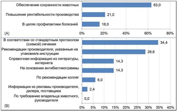
Results. It was found that 90.0% of the surveyed farm veterinarians kept an animal treatment log with records of antibiotic treatments, while 10.0% did not always record antibiotic prescriptions in the log. Of them, 63.0% use antibacterials to protect animals from diseases, 21.0% – to increase livestock profitability, and 16.0% – to prevent diseases. Most often, antibacterials were prescribed for treating diseases of the respiratory system (21.4%), the mammary gland (19.0%), and the reproductive organs (22.1%). More than half of the respondents stated that they do not use antibiotics for animal disease prevention, 17.3% use antimicrobials for preventing mammary gland diseases (mainly during the dry period), and 9.6% for preventing diseases of female reproductive organs after calving. Meanwhile, 50.0% of respondents do not aim to reduce their use of antibiotics. This creates high risks of the emergence of antibiotic-resistant strains of microorganisms and their spread through the food chain. The majority of veterinarians (68.0%) noted that the monitoring of residual concentrations of antibiotics in milk is a limiting factor for the widespread use of antibiotics in dairy cattle.
Conclusion. Despite the absence of a comprehensive program to reduce antibiotic use, the surveyed specialists acknowledge the existence of a mechanism that limits the spread of antimicrobial resistance. The only condition for curbing antibiotic use is the control of their content in raw milk, which dictates the need for further regulation in this area, as well as the optimization and prudent use of antibiotics.
ORIGINAL ARTICLES | BOVINE DISEASES
Introduction. Thelaziasis remains a widespread vector-borne parasitic zoonosis both within the Russian Federation and globally. Thelaziasis in yaks remains insufficiently studied, with the available data being fragmentary. It is the first time a thelaziasis clinical case in yaks from the Orenburg Oblast is described and it is of significant interest to a broad range of specialists.
Objective. Analysis and clinical case description of thelaziasis in yaks from the Orenburg Oblast, including the study of its causative agent and vector.
Materials and methods. Studies conducted from 2021 to 2023 at the steppe field station of the Institute of Steppe of the Ural Branch of the Russian Academy of Sciences in the Belyayevsky Raion of the Orenburg Oblast included clinical examinations and assessment of pathological lesions and severity of inflammation in the eyes and conjunctiva of yaks. Parasitic secretophagous dipterans (flies) from the ocular region were collected and counted, and their abundance, species and sex ratios were determined. Helminthoscopy was performed, and the nematode species was determined morphologically.
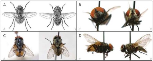
Results. Clinically, thelaziasis in yaks manifested as profuse lacrimation and recurrent keratoconjunctivitis. The extent of invasion (EI) was 100%, and the intensity of invasion (II) was 5. The detected helminths belonged to Thelazia rhodesi species. The intermediate hosts and vectors of Thelazia were facultative hematophages, specifically Musca autumnalis, a synbovine fly species ubiquitous in the steppe landscapes of the Orenburg Oblast. The ratio of females to males collected from the head region of yaks was 83 and 17%, respectively, confirming the leading role of female Musca autumnalis as vectors of nematodes of the genus Thelazia.
Conclusion. Domestic yaks in the natural and climatic conditions of the Orenburg Oblast are susceptible to thelaziasis. The disease progress, its clinical manifestations, as well as extent and intensity of invasion are likely influenced by acclimatization of yaks, who are not indigenous to this region.
Introduction. Cattle farming plays a key role in the agriculture of the Republic of Tajikistan, satisfying not only the needs for meat and dairy products. Recently, the intensification of livestock production in Tajikistan has been facing serious problems related to infectious diseases, in particular those caused by capripoxviruses, including lumpy skin disease (LSD), which causes significant economic damage and compromises animal performance and health in the region and around the world. Studying and understanding the LSD epizootology in the Republic of Tajikistan climatic environment will facilitate better disease control.
Objective. Epizootological data collection and brief description of LSD outbreaks in the Republic of Tajikistan in 2023.
Materials and methods. Traditional epizootological analysis tools were used to collect data on LSD outbreaks, as well as serological tools for the disease retrospective diagnosis.
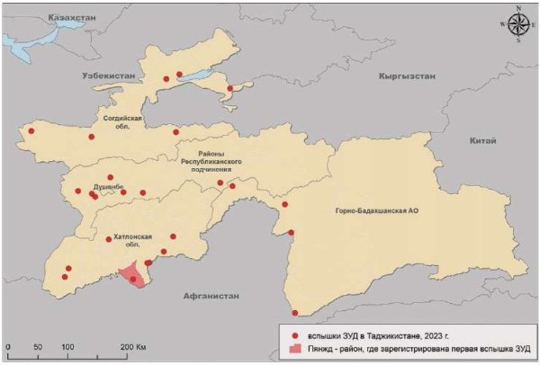
Results. In 2023, an LSD case was first reported in the Republic of Tajikistan in the region bordering Afghanistan. Analysis of the seasonal pattern of the disease occurrence in Tajikistan demonstrated that the LSD outbreaks were most often reported in summer and autumn: from July to November. The disease spread mainly in the areas with a high concentration of livestock, in particular in the Khatlon Region. The average animal density in the Khatlon Region is 46 cattle per 1 km2 and 117 sheep and goats per 1 km2. The peak of LSD epidemic was recorded in September – November 2023. Morbidity and mortality varied by districts, ranging from 10 to 55% and from 2 to 15%, respectively. Tests of 216 bovine serum samples demonstrated LSD virus antibodies in 109 animals, accounting for 50.5% of the total.
Conclusion. In 2023, a range of studies was conducted and measures were taken in the Republic of Tajikistan to prevent LSD spread. To effectively control the disease, it is necessary to strengthen epizootological monitoring, carry out timely vaccination of animals and implement measures for the identification and control of potential vectors. These steps will minimize economic losses and maintain the animal health in the republic.
ORIGINAL ARTICLES | PORCINE DISEASES
Introduction. Despite no cases of classical swine fever (CSF) have been recorded in the Russian Federation since 2021, gaining official recognition, as a disease-free zone, will require adoption of effective, safe vaccines compatible with the DIVA strategy. A range of expression systems is being evaluated as potential platforms for a recombinant subunit vaccine; synthesizing the E2 antigen in mammalian cells appears to be a particularly promising approach.
Objective. Optimizing transient transfection conditions in mammalian cell production lines for expression of classical swine fever virus (CSFV) E2 antigen.
Materials and methods. The nucleotide sequence encoding a 188-amino acid fragment of the E2 antigen was cloned into the pVAX1 vector. Transient transfection was performed using two common methods – calcium-phosphate and cationic (employing branched polyethylenimine, PEI) – on three established mammalian production cell lines: CHO-K1, PK-15, and BHK-21/13. Expression efficiency was controlled using immunofluorescence, quantitative reverse transcription polymerase chain reaction, and enzyme-linked immunosorbent assay.
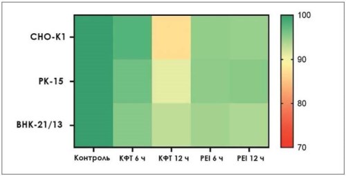
Results. It was determined that all the cell lines evaluated underwent transfection with an efficiency ranging from 60 to 90%. Cellular viability 24 hours post-transfection was at least 87%, with the lowest rates observed following calcium-phosphate transfection using an initial 12-hour incubation period. In all cases, transfection was accompanied by expression of specific messenger RNAs. The highest yield of the 17.3 kDa recombinant E2 protein was achieved in the CHO-K1 cell line (up to 47.4 mg/L), while the lowest yield was observed in the BHK-21/13 line (up to 24.1 mg/L). The specificity ratio in the antigen variant of the indirect enzyme-linked immunosorbent assay using specific antisera ranged from 5.1 to 6.2 units for all the expressed protein variants.
Conclusion. All the cell lines presented in the study demonstrated satisfactory transfection efficiency. Combined with their properties – such as high proliferation rates and adaptation to serum-free media – this makes them suitable for stable expression. Both the calcium-phosphate and cationic methods provide high transfection efficiency, relatively low cytotoxicity, and good reproducibility. The combined use of these control methods is advisable during the design phase of expression systems. In a production setting, however, the primary metric of their functionality is the overall yield of the specific recombinant protein, as determined by antigen-specific enzyme-linked immunosorbent assay.
Introduction. Classical swine fever (CSF) remains a critical challenge in global pig production. In the Russian Federation the last reported outbreak occurred in 2020 among wild boar populations, but the risk of re-emergence is sustained. To reduce the existing threats the targeted disease surveillance and control measures are needed to be improved.
Objective. To analyze the current classical swine fever situation and the outcomes of epizootic monitoring in the Russian Federation, and to develop evidence-based proposals for its improvement.
Materials and methods. This analysis draws upon laboratory test results from 2020 to 2024, as recorded in the “Vesta” electronic state information system (part of the FGIS “VetIS”); epidemiological data from the World Organization for Animal Health (WOAH); and the official “Guidelines for planning laboratory testing and sampling to improve classical swine fever surveillance in the Russian Federation”, developed and approved by the Federal Centre for Animal Health. Geospatial data were visualized using the MapChart platform, while statistical analyses were performed with Microsoft Excel.
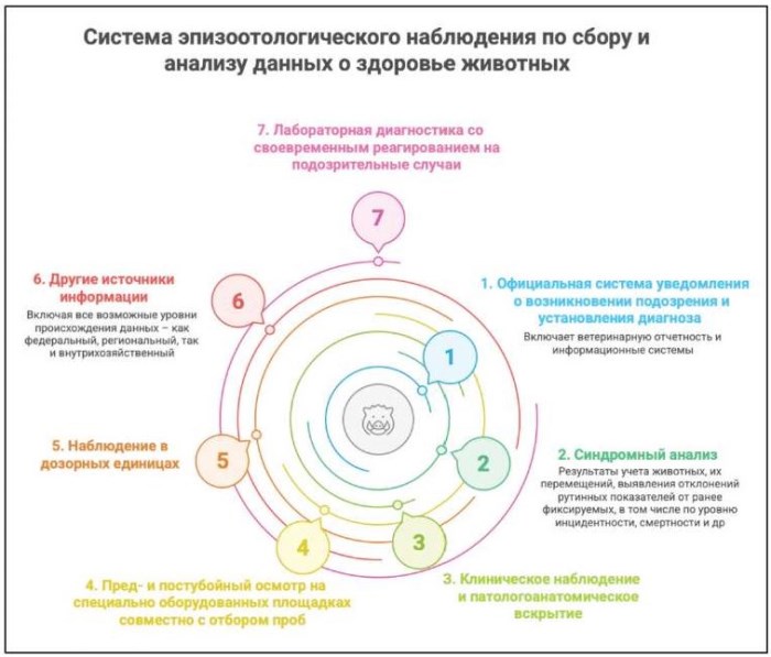
Results. Drawing on international experience in disease eradication and control, this study outlines a phased approach for the eradication of classical swine fever in the Russian Federation, with a view toward achieving official recognition of disease-free status from the World Organization for Animal Health. The proposed disease surveillance strategy is comprehensive and multifaceted, comprising: early detection measures, including immediate notification of suspected cases, syndromic analysis, and clinical examinations with necropsies; routine monitoring at key control points, such as ante-mortem and post-mortem inspections; and confirmatory procedures, consisting of strategic sampling, laboratory diagnostics, and surveillance in sentinel units. The study further explores the prospects for a strategic transition, including the zoning of Russian territory, the phased discontinuation of immunization with live (attenuated) vaccines, and the potential introduction of marker vaccines.
Conclusion. The proposed approaches are fully aligned with international standards and are specifically designed to achieve classical swine fever freedom in the Russian Federation. The full implementation of the proposed measures will significantly strengthen classical swine fever control in Russia and, consequently, enhance the export potential of the domestic pork industry.
ORIGINAL ARTICLES | AVIAN DISEASES
Introduction. Avian influenza virus (AIV) and Newcastle disease virus (NDV) pose serious threats to poultry health. Both pathogens are highly contagious and, due to their rapid spread, can lead to significant economic losses. Overlapping clinical signs complicate field differentiation of these diseases and delay response measures to isolate affected poultry. Rapid disease detection is critical for ensuring a timely response.
Objective. To develop a user-friendly test kit for detection of AIV and paramyxoviruses in organ samples from sick and dead chickens during disease outbreaks in commercial poultry operations.
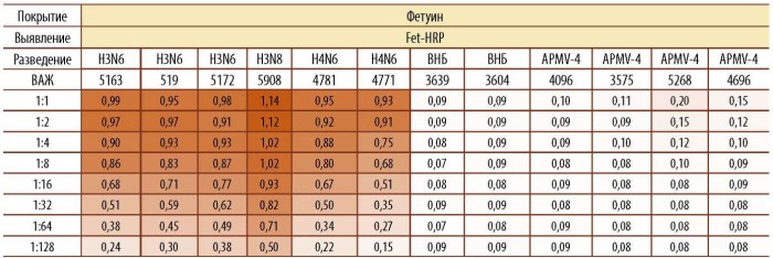
Materials and methods. To differentiate AIV and paramyxoviruses in pathological samples collected from sick and dead birds, 96-well plates coated with fetuin and anti-NDV IgY in designated wells were used. The results obtained were compared with those from polymerase chain reaction (PCR) and virus titration in chicken embryos.
Results. The developed method for pathogen detection is based on distinct virus-binding principles: influenza virus binds to a receptor analog, while paramyxoviruses bind to NDV specific antibodies. Previous studies using hundreds of strains have demonstrated that influenza A virus of various subtypes binds to the sialoglycosyl residues of bovine fetal serum protein – fetuin. In contrast, none of the paramyxovirus isolates tested bound to this sialoglycoprotein. For paramyxovirus capture, immunoglobulins isolated from the egg yolks of chickens immunized against NDV were utilized. Binding was performed in 96-well plates using a test-kit analogous to enzyme-linked immunosorbent assay (ELISA).
Conclusion. The developed method enables the identification and differentiation of AIV and NDV in organ tissue homogenates from infected chickens within a few hours, representing a significant step toward preventing the spread and facilitating the eradication of dangerous disease outbreaks.
ORIGINAL ARTICLES | BIOTECHNOLOGY
Introduction. Chitosan immunomodulatory and mucoadhesive properties render it a promising vaccine adjuvant. Safety – particularly the absence of cytotoxicity – is a key requirement for adjuvant candidates. In vitro biocompatibility assessments enable evaluation of chitosan preparations prior to animal testing.
Objective. To evaluate low molecular weight chitosan solution at a concentration of 10 mg/mL for its cytotoxic effect on chicken embryo fibroblast (CEF) cultures and calf coronary artery epithelial-like cells (CCEC) to justify its further use as a vaccine adjuvant.
Materials and methods. Low molecular weight (LMW) chitosan (degree of deacetylation: 90%) prepared with a 1% glutamic acid solution (pH 6.9) was used. Cytotoxicity was comprehensively assessed using three methods: trypan blue vital staining (for cell viability), live-cell microscopy (for morphological evaluation), and calculation of the proliferation index after 72 hours of incubation at 37 °C in a 5% CO2 atmosphere.
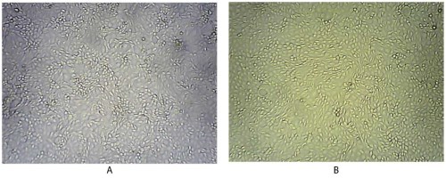
Results and discussion. Following 2-hour incubation with chitosan, viable CEF and CCEC were 97.4 and 98.7%, respectively, with no significant differences from controls (97.6 and 96.4%). Microscopy at 72 hours showed dense, homogeneous monolayers in test groups, free of cytopathic effects, vacuolization, or morphollogical changes – indistinguishable from controls. Proliferation indices aligned closely (CEF: 3.9 and 3.6; CCEC: 3.7 and 3.8), evidencing no cytostatic effect of the chitosan preparation.
Conclusion. Low-molecular-weight chitosan (10 mg/mL) exhibited no in vitro cytotoxic or cytostatic effects on the tested cell lines. The findings confirm its biocompatibility and justify advancement to in vivo studies for developing safe, effective vaccines for veterinary use.
ANNIVERSARY DATES




ISSN 2658-6959 (Online)


