



Contents
Scroll to:
https://doi.org/10.29326/2304-196X-2026-15-1-38-45
Scroll to:
Introduction. Listeria monocytogenes is a pathogenic microorganism that causes a large number of deaths due to the consumption of contaminated animal products. This underpins the relevance of monitoring the spread of the listeriosis agent in raw materials, animal products, and environmental objects, as well as the antibiotic resistance of isolates.
Objective. Serotype identification and antibiotic resistance analysis of Listeria monocytogenes isolates recovered from animal products in 2021–2024.
Materials and methods. The work was performed at the Microbiology Unit of the Vladimir Testing Laboratory, the Federal Centre for Animal Health. Listeria genus bacteria isolates were identified using time-of-flight mass spectrometry. Antibiotic resistance of isolates belonging to the species Listeria monocytogenes was determined by the disk diffusion method. The values of the growth retardation zones were interpreted according to the Russian recommendations “Determination of the sensitivity of microorganisms to antimicrobial drugs” (IACMAC, version 2025-01). Listeria serological groups were identified using real-time polymerase chain reaction (qPCR) with primers manufactured by Syntol (Russia).
Results. The article presents the results of antibiotic resistance testing of 77 Listeria monocytogenes isolates detected in animal products in 2021–2024, as well as their differentiation by serogroups. Listeria monocytogenes was most frequently detected in poultry products. The detected isolates showed maximum resistance to cefuroxime, sulfamethoxazole/trimethoprim, norfloxacin, rifampicin, levofloxacin, and kanamycin. Moreover, most isolates exhibited resistance to more than one antimicrobial medicinal product. The study established the belonging of the Listeria monocytogenes isolates to the following serogroups: IIa (serotypes 1/2a, 3a) – 92.2%; IIc (serotypes 1/2c, 3c) – 5.2%; IVb (serotypes 4b, 4d, 4e) – 2.6%.
Conclusion. The spread of resistance, including multidrug resistance, among Listeria monocytogenes isolates detected in animal products in 2021–2024 was demonstrated. The study identified the presence of listeria belonging to group IVb (serotypes 4b, 4d, 4e). However, the dominant part of Listeria monocytogenes isolates was classified as group IIa (serotypes 1/2a, 3a).
Akulich O.А., Shadrova N.B., Denisova G.S. Serotype identification and antibiotic resistance analysis of Listeria monocytogenes isolates recovered from animal products in 2021–2024. Veterinary Science Today. 2026;15(1):38-45. https://doi.org/10.29326/2304-196X-2026-15-1-38-45
Listeria monocytogenes is a gram-positive motile facultative anaerobic rod that does not form spores or capsules and causes listeriosis, a life-threatening disease manifesting, other things, as central nervous system damage and meningoencephalitis. Children, including newborns, pregnant women (leading to fetal death), and the elderly are at high risk. In healthy individuals, L. monocytogenes causes mild symptoms, but listeriosis poses a serious threat to immunocompromised individuals and those with chronic diseases, as it can lead to sepsis and other complications [1][2][3][4].
This microorganism is widespread in terrestrial and aquatic ecosystems, being transmitted to animals and humans both directly and indirectly through the consumption of contaminated food or water. L. monocytogenes infection mainly characterized by foodborne transmission, and according to the World Health Organization, listeriosis is one of the most serious and severe foodborne diseases, with the highest case fatality rates in foodborne infections (over 20%) [5][6][7].
L. monocytogenes bacteria can adapt to various conditions: they can survive at temperatures below 7 °C. They are resistant to low pH values and high salt concentrations. Furthermore, Listeria can form biofilms, that allow them not only to persist in food production environments, but also to accumulate, even if their initial concentration was low. Animal products with a high risk of L. monocytogenes contamination include meat preparations and ready-to-eat meat products, including vacuum-packed products, dairy products, including soft cheeses, and cold-smoked fish products. Chilled ready-to-eat foods pose a particular risk [8][9][10].
The disease is reported in more than 65 countries on all continents. The number of recorded cases per year is 0.1–10.0 per 1 million people, depending on the country [9][11].
In the European Union and European Economic Area (EU/EEA) countries, 2,993 confirmed cases were reported in 2023 (0.67 cases per 100,000 population), the highest annual rate since 2007. Incidence peaks during the summer months, with the number of cases increasing annually, showing a statistically significant rise and upward trend [12][13].
Analysis of listeriosis epidemiology demonstrates that most sporadic cases and all major epidemic outbreaks are associated with pathogen isolates belonging to the first two phylogenetic lineages, designated I and II. In total, four phylogenetic lineages are distinguished within the species L. monocytogenes, differing in genetic and phenotypic characteristics.
Traditionally, five serogroups are distinguished: I (serotypes 1/2a and 3a; with 3a rarely detected in clinical cases); II (serotypes 1/2b, 3b and 7); III (serotypes 1/2c, 3c; with 3c being an extremely rare form); IV (serotypes 4b, 4d and 4e; with 4e playing a minor role in disease development); V (serotypes 4ab, 4a, 4c, which are practically not associated with clinical cases).
Listeria serotypes, classified based on variations in somatic (O) and flagellar (H) antigens, show significant differences in their epidemic potential and pathogenicity. These differences are due to the heterogeneity of antigenic structure, allowing serotypes to adapt to different environmental conditions and interact with the host’s immune system [3][5][6][14][15].
Based on molecular typing, L. monocytogenes is divided into the following serogroups: IIa (serotypes 1/2a and 3a), IIb (serotypes 1/2b, 3b), IIc (serotypes 1/2c, 3c and 7), and IVb (serotypes 4b, 4d and 4e) [12][16].
Human listeriosis is most commonly caused by serotypes 1/2a, 1/2b, and 4b, which account for over 90% of detected isolates. Serotype 4b is associated with most major listeriosis outbreaks, making it one of the most dangerous. Meanwhile, the vast majority of isolates belonging to serotypes 1/2 are widely distributed in food products and ecological niches where Listeria spp. are found. In particular, serotype 1/2a is most often detected in food products [6][17][18][19].
In the Russian Federation, the number of listeriosis cases and the percentage of fatalities are also increasing annually (an increase in severe and moderate forms of the disease is noted), despite the fact that listeriosis is generally reported as sporadic cases [2][10].
Thus, according to the official reports of the Federal Service for the Oversight of Consumer Protection and Welfare, in 2021, the incidence of listeriosis in the Russian Federation was 45 cases1; in 2022 – 81 cases, including 14 fatalities2; in 2023 – 100 cases, including 18 fatalities3; in 2024 – 208 cases, 49 of which were fatal4. The majority of cases are reported annually in large cities, Moscow and Saint Petersburg, with no pronounced seasonal fluctuations of the disease established [1].
The high mortality rate of listeriosis requires prompt treatment, specifically the use of antibiotics. Regarding antimicrobial susceptibility, L. monocytogenes, despite its widespread distribution in the environment, generally exhibits relatively low resistance rates. However, recent studies have shown the acquisition of antibiotic resistance in Listeria strains, including those isolated from food products [3][16][20][21][22].
At the same time, antibiotic resistance is currently considered one of the main threats to global health, to combat which the World Health Organization, the Food and Agriculture Organization of the United Nations, the United Nations Environment Programme, and the World Organisation for Animal Health have united under the “One Health” concept [23][24][25].
To combat antimicrobial resistance in the Russian Federation, the “Strategy for Preventing the Spread of Antimicrobial Resistance in the Russian Federation for the Period up to 2030”5 was approved in 2017. Furthermore, in 2024, an “Action Plan for 2025–2030”6 for the implementation of this strategy was approved, determining legal regulation, public awareness, systematic monitoring, and more.
Thus, the relevance of this study stems from the importance of monitoring the spread of L. monocytogenes by testing food products to trace the epidemiological pathways of the pathogen, including resistant ones, prevent its transmission to humans, and occurrence of listeriosis outbreaks.
The novelty of the work lies in the results of tested samples of livestock products obtained from three regions of Central Russia (Vladimir, Kostroma, and Ivanovo Oblasts), with subsequent isolation of L. monocytogenes isolates, typing using qPCR, determination of antibiotic resistance, and evaluation of trends in antibiotic resistance.
The objective of the work is to identify serotypes and analyze the antibiotic resistance of L. monocytogenes isolates recovered from animal products between 2021 and 2024.
The testing was conducted in the Microbiological Testing of the Vladimir Testing Laboratory of the Federal Centre for Animal Health. A total of 77 L. monocytogenes isolates, recovered from animal products during 2021–2024, were used in this testing.
Reagents and nutrient media: Fraser broth for primary enrichment (Merck KGaA, Germany), Fraser broth for secondary enrichment (Merck KGaA, Germany), Ottaviani – Agosti agar (ALOA; Merck KGaA, Germany), Oxford agar (Merck KGaA, Germany), tryptone soya agar (TSA; State Research Centre for Applied Microbiology and Biotechnology, Russia), and Mueller – Hinton agar (MHA; State Research Centre for Applied Microbiology and Biotechnology, Russia).
Microbiological testing was performed according to GOST 32031-2022 “Food products. Methods for detection of Listeria monocytogenes and other species of Listeria (Listeria spp.)”7.
A 25 g test sample of the product was placed into a sterile bag containing 225 mL of Fraser broth for primary enrichment, homogenized for 1 min, and incubated at (30 ± 1) °C for (25 ± 1) hours. After primary enrichment, 0.1 mL of the culture was transferred into 10 mL of Fraser broth and incubated at (37 ± 1) °C for (24 ± 2) hours.
Following incubation, samples subcultured using a bacteriological loop onto the surface of two solid selective media (ALOA and Oxford agar) and cultured at (37 ± 1) °C for 24–48 hours, monitoring for the presence of colonies characteristic of Listeria spp.
Colonies exhibiting growth characteristic of Listeria spp. were subcultured onto the surface of tryptone soya agar with yeast extract to obtain isolated colonies and incubated at (37 ± 1) °C for (24 ± 3) hours.
Simultaneously, rapid identification of the isolated microorganisms was performed using time-of-flight mass spectrometry (Autof MS1000, Autobio Diagnostics Co., Ltd, China), as well as by determining culture motility, Gram stain, and catalase activity.
Determination of antibiotic resistance. Antimicrobial susceptibility testing of L. monocytogenes isolates was performed using the disk diffusion method according to the methodological guidelines MUK 4.2.1890-04 “Determination of the Sensitivity of Microorganisms to Antimicrobials”8.
The following antibiotics (paper disks manufactured by the Saint-Petersburg Pasteur Institute, Russia) were used: azithromycin (15 µg), amikacin (30 µg), amoxicillin (20 µg), ampicillin/sulbactam (10 µg), benzylpenicillin (10 IU/6 µg), vancomycin (30 µg), doxycycline (30 µg), imipenem (10 µg), kanamycin (30 µg), levofloxacin (5 µg), meropenem (10 µg), norfloxacin (10 µg), rifampicin (5 µg), sulfamethoxazole/trimethoprim (23.75/1.25 µg), streptomycin (10 µg), tetracycline (30 µg), chloramphenicol (30 µg), cefazolin (30 µg), cefuroxime (30 µg), and erythromycin (15 µg).
The choice of antibacterial agents was driven by the fact that penicillins (ampicillin, benzylpenicillin, penicillin, amoxicillin) are often used in combination with aminoglycosides (gentamicin, streptomycin) in therapy of animals, as well as in the treatment of listeriosis in humans. Alternative antibiotics (second-line treatment agents) may include: sulfamethoxazole/trimethoprim, macrolides (erythromycin), fluoroquinolones (levofloxacin), tetracyclines (tetracycline, doxycycline), carbapenems (meropenem, imipenem), rifampicin, and vancomycin. Thus, detection of resistance to these agents may limit treatment options, particularly for patients with allergic reactions to certain antimicrobials [5][16][21][22][26].
For antibiotic susceptibility testing, a bacterial suspension with an optical density of 0.5 McFarland standard was prepared from a 24-hour culture of L. monocytogenes isolates grown on MHA.
The density of the suspension was measured using a densitometer (VITEK® bioMérieux model Densichek, France). Subsequently, it was inoculated onto sterile Petri plates (on dried surface of tryptone soya agar) with a sterile cotton swab by streaking with no gaps. After applying antibiotic disks (4 disks per Petri plate), the plates were incubated at 37 °C for (18 ± 2) hours. The growth retardation zones of microorganisms around the discs were measured with an accuracy of 1 mm.
Results were interpreted using the Russian guidelines “Determination of the Sensitivity of Microorganisms to Antimicrobials” (IACMAC, version 2025-01), prepared based on the recommendations of the European Committee on Antimicrobial Susceptibility Testing (EUCAST) [27][28].
Since EUCAST recommendations do not provide criteria for interpretation of L. monocytogenes antibiotic resistance for the entire list of antimicrobials used in this work, the zone diameter breakpoints for most antibiotics were based on data for Staphylococcus spp. For the analysis of Listeria susceptibility to vancomycin and streptomycin, values for Enterococcus spp. were used [5][16][29][30].
Real-time PCR (qPCR). For DNA extraction, the “RIBO-prep” reagent kit (Central Research Institute of Epidemiology of Rospotrebnadzor, Russia) was used according to the manufacturer’s instruction.
Serological groups of L. monocytogenes were identified according to the methodological recommendations for the differentiation of bacterial genome regions of serogroups (1/2a, 3a), (1/2c, 3c), and (4b, 4d, 4e) in animal products using qPCR, developed at the Federal Centre for Animal Health.
Serogroup IIa (serotypes 1/2a and 3a) was identified by amplification of the lmo0737 gene fragment; the lmo0737 and lmo1118 genes allowed identification of serogroup IIc (serotypes 1/2c, 3c); serogroup IVb (serotypes 4b, 4d, and 4e) was determined by amplification of the ORF0799 gene. Primers were manufactured by order of Syntol (Russia).
The following strains were used as positive controls:
– serotype 1/2a – DNA of L. monocytogenes No. 15 (Federal Research Center for Virology and Microbiology, Russia);
– serotype 1/2c – DNA of L. monocytogenes 5348 No. 20 (Federal Research Center for Virology and Microbiology);
– serotype 3a – DNA of L. monocytogenes No. 39 (Federal Research Center for Virology and Microbiology);
– serotype 3c – DNA of L. monocytogenes No. 46 (Federal Research Center for Virology and Microbiology);
– serotype 4b – DNA of L. monocytogenes ATCC 19115 (State Research Center for Applied Microbiology and Biotechnology, Russia);
– serotype 4d – DNA of L. monocytogenes 10888 No. 72 (Federal Research Center for Virology and Microbiology);
– serotype 4e – DNA of L. monocytogenes 19118 No. 75 (Federal Research Center for Virology and Microbiology).
The nucleotide sequences of primers and probes for differentiating genomic regions of L. monocytogenes serogroups (1/2a, 3a), (1/2c, 3c), and (4b, 4d, 4e) are presented in the Table [14][19].
Table
Primers for the molecular identification and differentiation of L. monocytogenes isolates by serogroups
Serogroups | Genes | Sequence (5’–3’) |
4b, 4d, 4e | ORF0799F | 5’-GCTGGGTTTCTTACGA-3’ |
ORF0799R | 5’-CAACCGTTCATTTAGCTCAT-3’ | |
ORF0799P | FAM-TCTGCTGTTCAGTTATTGGAGTGGGA-BHQ1 | |
1/2а, 1/2c, 3а, 3с | Lmo0737F | 5’-GCGGATGTGATTGATTTAC-3’ |
Lmo0737R | 5’-AAACTGCACTAACTCTTGAAT-3’ | |
Lmo0737P | ROX -TGCTCCAGGATCAAGACACGGTA-BHQ2 | |
1/2c, 3c | Lmo1118F | 5’-CTTAGTATTCCAGGATTTAAGACC-3’ |
Lmo1118R | 5’-CCAAAGAACCAAATTGATCGAATC-3’ | |
Lmo1118P | FAM-CCTTTATCTTCTCCTGAGTGTATACGCCTC-RTQ1 |
When preparing the reaction mixture, the following volumes of components per sample were used: 10× PCR buffer B – 2.5 µL; dNTP 2.5 mM – 2.5 µL; MgCl2 25 mM – 2.5 µL; a mixture of primers and a probe (10 pmol/µL each) – 0.5 µL each; SynTaq DNA polymerase 5 U/µL – 0.2 µL; ddH2O – 11.8 µL (a set of reagents for qPCR manufactured by Syntol, Russia).
The qPCR was performed in a thermal cycler (CFX module, C1000 Touch, Bio-Rad Laboratories, Inc., USA) in a volume of 25 µL containing 20 µL of a mixture and 5 µL of L. monocytogenes DNA isolates.
The mixture preparation protocol included heating of the reaction mixture at 94 °C for 3 minutes, 40 cycles with denaturation at 94 °C for 20 seconds, annealing at 58 °C for 30 seconds and elongation at 72 °C for 25 seconds, and then completion of the reaction at 72 °C for 10 minutes.
Statistical processing of the research results was performed using the Microsoft Excel program.
During testing of samples of animal products from 2021 to 2024, a total of 77 isolates of L. monocytogenes were detected (12 isolates in 2021, 22 isolates in 2022, 22 isolates in 2023, 21 isolates in 2024).
Figure 1 presents a graphical interpretation of the distribution frequency of L. monocytogenes isolates detected in animal products. The pathogen was most frequently found in poultry meat, with 48 Listeria isolates, accounting for a significant proportion (62%) of the total detected isolates. Fish and fish products, as well as beef and meat preparations derived from it, also represented a significant source of L. monocytogenes contamination (16 isolates – 21% and 10 isolates – 13%, respectively).
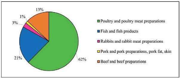
Fig. 1. Distribution frequency of L. monocytogenes isolates detected in animal product samples in 2021–2024
Data from the European Food Safety Authority (EFSA) indicate that outbreaks of foodborne infections caused by L. monocytogenes in Europe were mainly attributed to contamination of food products from these same categories: broiler meat, beef, pork, and products thereof; fish and fishery products; and cheeses [7].
As part of this work, evaluation of the resistance of L. monocytogenes isolates to 20 antimicrobial agents was conducted. The results are presented in Figure 2.
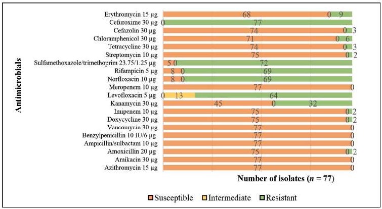
Fig. 2. Antibiotic resistance of L. monocytogenes isolates detected in animal products in 2021–2024
The research demonstrated a high level of resistance in L. monocytogenes isolates to several antibacterials. Thus, the maximum frequency of resistance was observed to cefuroxime (100.0%), sulfamethoxazole/trimethoprim (93.5%), norfloxacin (89.6%), rifampicin (89.6%), levofloxacin (83.1%), kanamycin (41.6%). Concurrently, all L. monocytogenes isolates were susceptible to ampicillin/sulbactam, benzylpenicillin, azithromycin, amikacin, vancomycin, and meropenem.
The data obtained correlate with findings of other authors that show the susceptibility of L. monocytogenes isolates to ampicillin, benzylpenicillin, vancomycin and resistance to rifampicin, sulfamethoxazole/trimethoprim, kanamycin, norfloxacin and erythromycin [4][5][22][31]. Furthermore, the Federal Service for Surveillance on Consumer Rights Protection and Human Wellbeing, in its official report for 2024, provided information on the resistance of L. monocytogenes strains isolated from food products to sulfamethoxazole/trimethoprim9.
These and other results presented by individual authors emphasize the importance of monitoring the susceptibility of L. monocytogenes due to the increasing resistance to a number of antimicrobials, including tetracycline and erythromycin [3]. In our work, 3.9% of isolates were found to be resistant to tetracycline, and 11.7% to erythromycin.
Additionally, several researchers note the diversity of genetic profiles of L. monocytogenes, which leads to variability in antimicrobial susceptibility [16].
In the current tests, it was found that 98.7% of L. monocytogenes isolates were resistant to more than one antibiotic. No isolates resistant to all tested antimicrobials were detected.
Results from other authors not only confirm the fact of resistance of L. monocytogenes isolated from food products to at least one antibiotic, but also demonstrate an increase in the number of strains with multidrug resistance, posing a serious challenge for modern medicine [5][16][32].
Furthermore, as shown in Figure 3, resistance to five antimicrobials was identified in 30 isolates (38.9%), to six in 18 isolates (23.4%), and to four in 12 isolates (15.6%).

Fig. 3. L. monocytogenes isolates with multidrug resistance detected in samples of animal products in 2021–2024
Also, as part of the work, 2 L. monocytogenes isolates resistant to eight antibiotics (2.6%) and 12 isolates resistant to seven antibiotics (15.6%) were identified.
Nearly half of the isolated L. monocytogenes strains (46.8%) were resistant to three classes of antibiotics (cephalosporins, sulfonamides, fluoroquinolones) and rifampicin. Isolates with multidrug resistance were most frequently detected in poultry meat products.
The emergence of strains resistant to fluoroquinolones was first reported in the early 1990s; however, multidrug resistance in L. monocytogenes was rare until the mid-2000s [5].
Figure 4 demonstrates the increase in the number of L. monocytogenes isolates resistant to certain antimicrobials within a single class (fluoroquinolones) during the period from 2021 to 2024. Specifically, in 2024, isolate resistance to levofloxacin (a third-generation fluoroquinolone) increased by 11.9% compared to 2021. Similar results were observed for norfloxacin (a second-generation fluoroquinolone), with a 20.2% increase in resistance over the four-year period. Similar results were observed for norfloxacin (a second-generation fluoroquinolone), with a 20.2% increase in resistance over the four-year period.
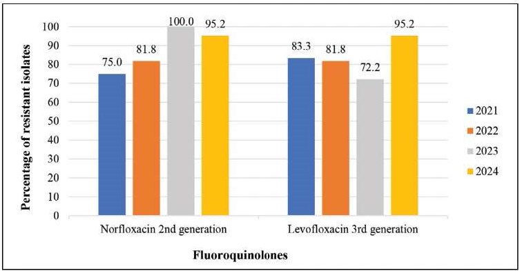
Fig. 4. Resistance of L. monocytogenes isolates to fluoroquinolones
Resistance to kanamycin (a first-generation aminoglycoside) increased by 5.9% between 2021 and 2024. A high level (ranging from 81.8% to 100.0%) of resistance to sulfamethoxazole/trimethoprim and rifampicin was noted (Fig. 5).

Fig. 5. Resistance of L. monocytogenes isolates to sulfamethoxazole/trimethoprim, rifampicin, and kanamycin
In the next stage of our work, the serological groups of L. monocytogenes were identified. According to findings from other authors, a significant proportion of Listeria isolates detected in products belong to serogroup IIa, particularly serotype 1/2a, which demonstrates higher adaptability and resistance to disinfectants or other environmental factors [3][6][16][32][33]. However, according to data from the European Centre for Disease Prevention and Control (ECDC European Surveillance System, TESSy) report, in 2023, the most prevalent serogroup was IVb (47.8%), followed by IIa (41.7%), IIb (9.0%), and IIc (1.6%) [12].
Determining Listeria serotypes using traditional serological methods is time-consuming, lacks specificity, and is not widespread in the Russian Federation due to the absence of specific sera. Several authors recommend using qPCR for determining L. monocytogenes serogroups [15][17].
In our work, using qPCR with three pairs of primers for the serological identification of 77 L. monocytogenes isolates, it was determined that 71 isolates (92.2%) belonged to serotypes 1/2a, 3a and were assigned to serogroup IIa; 4 isolates (5.2%) belonged to serotypes 1/2c, 3c and serogroup IIc; and 2 isolates (2.6%) belonged to serotypes 4b, 4d, 4e and serogroup IVb (Fig. 6). An isolate of the most dangerous serotype, L. monocytogenes 4b, was detected in poultry meat, which may pose a potential epidemiological hazard.

Fig. 6. Determination of L. monocytogenes serotypes in animal product samples by qPCR
In this work, 77 L. monocytogenes isolates were identified, and it was found that poultry meat was the main source of Listeria contamination, accounting for 62% of the total isolates detected.
Data were obtained indicating increasing resistance in Listeria isolates, including multidrug resistance.
The L. monocytogenes isolates exhibited the highest resistance rates to cefuroxime (100.0%), sulfamethoxazole/trimethoprim (93.5%), norfloxacin (89.6%), rifampicin (89.6%), levofloxacin (83.1%), and kanamycin (41.6%). At the same time, all isolates were susceptible to azithromycin, amikacin, ampicillin/sulbactam, benzylpenicillin, vancomycin, and meropenem.
The vast majority of L. monocytogenes isolates (98.7%) demonstrated resistance to more than one antibiotic. Thus, resistance to five antimicrobials was observed in 30 isolates (38.9%), to six in 18 isolates (23.4%), and to four in 12 isolates (15.6%). Additionally, 2 isolates (2.6%) were found to be resistant to eight antibiotics, and 12 isolates (15.6%) to seven antibacterials.
Analysis of the data obtained from 2021 to 2024 revealed an increase in resistance in L. monocytogenes isolates to medicines from the fluoroquinolone group: resistance to norfloxacin (second-generation fluoroquinolone) increased by 20.2%, and to levofloxacin (third-generation fluoroquinolone) by 11.9%. During the same period, an increase in resistance to kanamycin by 5.9% was also observed. Resistance to sulfamethoxazole/trimethoprim and rifampicin remained at levels between 81.8 and 100.0%.
Using qPCR, it was determined that 92.2% of the studied L. monocytogenes isolates belong to serogroup (1/2a, 3a). Isolates belonging to serogroup IVb, which includes the most epidemiologically dangerous Listeria serotype 4b, were detected in poultry meat.
Contribution of the authors: Akulich O. А. – testing, obtained data analysis and interpretation, paper text preparation, figure creation; Shadrova N. B. – formulation of key goals and objectives, paper text editing and approval of final paper text; Denisova G. S. – conceptualization, formulation of key goals and objectives, paper text editing.
Вклад авторов: Акулич О. А. – проведение исследований, анализ и интерпретация полученных данных, подготовка текста статьи, создание рисунков; Шадрова Н. Б. – формулировка ключевых целей и задач, редактирование текста статьи и утверждение окончательного варианта; Денисова Г. С. – формирование идеи, формулировка ключевых целей и задач, редактирование текста статьи.
1. https://www.rospotrebnadzor.ru/documents/details.php?ELEMENT_ID=21796 (in Russ.)
2. https://www.rospotrebnadzor.ru/documents/details.php?ELEMENT_ID=25076 (in Russ.)
3. https://www.rospotrebnadzor.ru/documents/details.php?ELEMENT_ID=27779 (in Russ.)
4. https://www.rospotrebnadzor.ru/documents/details.php?ELEMENT_ID=30171 (in Russ.)
5. http://static.government.ru/media/files/onJ3GY3ObDGqLDvrED7AhpLF3ywRRFpp.pdf (in Russ.)
6. https://www.garant.ru/products/ipo/prime/doc/409448585/?ysclid=mhymceuaxf332408554 (in Russ.)
7. https://docs.cntd.ru/document/1200193714?ysclid=mhyosx40nk732110910 (in Russ.)
8. https://docs.cntd.ru/document/1200038583?ysclid=mhypcum520443253115 (in Russ.)
9. https://www.garant.ru/products/ipo/prime/doc/409448585/?ysclid=mhymceuaxf332408554 (in Russ.)
1. Kovalev V. A., Filatov N. N., Aleshina E. N., Simonova E. G. Sickness of listeriosis in Russian Federation. Science of the Young (Eruditio Juvenium). 2019; 7 (4): 509–517. https://doi.org/10.23888/HMJ201974509-517
2. Chestnova T. V., Malyutina T. K., Gusakova D. R., Zaytseva E. D. Dynamics of the epidemic process at listeriosis in Russia and the world (literature review). Journal of New Medical Technologies. 2024; (6): 101–110. https://doi.org/10.24412/2075-4094-2024-6-2-1 (in Russ.)
3. Mikhailova Yu. V., Molchanov A. D., Shelenkov A. A., Tyumentseva M. A., Karbyshev K. S., Tyumentsev A. I., et al. Heterogeneity of antibiotic- resistant isolates of Listeria monocytogenes isolated from food products in Moscow. Epidemiology and Vaccinal Prevention. 2023; 22 (6): 108–123. https://doi.org/10.31631/2073-3046-2023-22-6-108-123 (in Russ.)
4. Shastin P. N., Yakimova E. A., Supova A. V., Savinov V. A., Ezhova E. G., Khabarova A. V., Laishevtsev A. I. Antibiotic resistance and phage sensitivity of topical listeriosis pathogens. Agrarian science. 2024; (3): 50–56. https://doi.org/10.32634/0869-8155-2024-380-3-50-56 (in Russ.)
5. Ermolaeva S. A., Karpova T. I., Andriyanov P. A., Zhurilov P. A., Voronina O. L., Ryzhova N. N., et al. Distribution of antimicrobial resistance among clinical and food Listeria monocytogenes isolated in Moscow in 2019–2021. Clinical Microbiology and Antimicrobial Chemotherapy. 2022; 24 (2): 156–164. https://doi.org/10.36488/cmac.2022.2.156-164 (in Russ.)
6. Astashkin E. I., Alekseeva E. A., Borzenkov V. N., Kislichkina A. A., Mukhina T. N., Platonov M. E., et al. Molecular genetic characteristics of polyresistant Listeria monocytogenes strains and identification of new sequence types. Molecular Genetics, Microbiology and Virology. 2021; 36 (4): 159–169. https://doi.org/10.3103/S0891416821040029
7. EFSA (European Food Safety Authority). Story map on Listeria monocytogenes. 2024. https://storymaps.arcgis.com/stories/629e6627e6c64111bfd-5b9257473c74a
8. Churchill K. J., Sargeant J. M., Farber J. M., O’Connor A. M. Prevalence of Listeria monocytogenes in select ready-to-eat foods – deli meat, soft cheese, and packaged salad: A systematic review and meta-analysis. Journal of Food Protection. 2019; 82 (2): 344–357. https://doi.org/10.4315/0362-028x.jfp-18-158
9. World Health Organization. Listeriosis. https://www.who.int/news-room/fact-sheets/detail/listeriosis
10. Voronina O. L., Kunda M. S., Ryzhova N. N., Kutuzova A. V., Akse nova E. I., Karpova T. I., et al. Listeriosis: genotyping as a key for identification a possible source of infection. Clinical Microbiology and Antimicrobial Chemotherapy. 2019; 21 (4): 261–273. https://doi.org/10.36488/cmac.2019.4.261-273 (in Russ.)
11. Tyukavkina S. Yu., Kotieva I. M., Dodokhova M. A., Grechina D. A., Babiev S. A., Kharseeva G. G. Pathogenesis and clinical forms of human listeriosis. South Russian Journal of Therapeutic Practice. 2024; 5 (1): 99–111. https://doi.org/10.21886/2712-8156-2024-5-1-99-111 (in Russ.)
12. European Centre for Disease Prevention and Control. Listeriosis. In: ECDC. Annual Epidemiological Report for 2023. Stockholm: ECDC; 2025. https://www.ecdc.europa.eu/sites/default/files/documents/LIST_AER_2023_Report.pdf
13. European Food Safety Authority (EFSA), European Centre for Disease Prevention and Control (ECDC). The European Union One Health 2023 Zoonoses report. EFSA Journal. 2024; 22 (12):e9106. https://doi.org/10.2903/j.efsa.2024.9106
14. Vitullo M., Grant K. A., Sammarco M. L., Tamburro M., Ripabelli G., Amar C. F. L. Real-time PCRs assay for serogrouping Listeria monocytogenes and differentiation from other Listeria spp. Molecular and Cellular Probes. 2013; 27 (1): 68–70. https://doi.org/10.1016/j.mcp.2012.10.001
15. Alía A., Andrade M. J., Córdoba J. J., Martín I., Rodríguez A. Development of a multiplex real-time PCR to differentiate the four major Listeria monocytogenes serotypes in isolates from meat processing plants. Food Microbiology. 2020; 87:103367. https://doi.org/10.1016/j.fm.2019.103367
16. Tsitsos A., Peratikos P., Damianos A., Kyritsi M. A., Arsenos G., Hadjichristodoulou C., et al. Prevalence, molecular characterization, antibiotic resistance, and investigation of transmission pathways of Listeria monocytogenes strains isolated along the beef production chain. Food Microbiology. 2025; 129:104745. https://doi.org/10.1016/j.fm.2025.104745
17. Khaptanova N. M., Аndreevskaya N. M., Lukyanova S. V., Konovalova Zh. A., Gefan N. G., Ostyak А. S., Tokmakova E. G. Aspects of serological diagnostics of listeriosis (literature review). Acta Biomedica Scientifica. 2019; 4 (1): 43–49. https://doi.org/10.29413/ABS.2019-4.1.7 (in Russ.)
18. Buchanan R. L., Gorris L. G. M., Hayman M. M., Jackson T. C., Whiting R. C. A review of Listeria monocytogenes: An update on outbreaks, virulence, dose-response, ecology, and risk assessments. Food Control. 2017; 75: 1–13. https://doi.org/10.1016/j.foodcont.2016.12.016
19. Pan Y., Breidt F. Jr., Kathariou S. Competition of Listeria monocytogenes serotype 1/2a and 4b strains in mixed-culture biofilms. Applied and Environmental Microbiology. 2009; 75 (18): 5846–5852. https://doi.org/10.1128/AEM.00816-09
20. Olaimat A. N., Al-Holy M. A., Shahbaz H. M., Al-Nabulsi A. A., Abu Ghoush M. H., Osaili T. M., et al. Emergence of antibiotic resistance in Listeria monocytogenes isolated from food products: A comprehensive review. Comprehensive Reviews in Food Science and Food Safety. 2018; 17 (5): 1277–1292. https://doi.org/10.1111/1541-4337.12387
21. Radoshevich L., Cossart P. Listeria monocytogenes: towards a complete picture of its physiology and pathogenesis. Nature Reviews Microbio logy. 2018; 16 (1): 32–46. https://doi.org/10.1038/nrmicro.2017.126
22. Baquero F., Lanza V. F., Duval M., Coque T. M. Ecogenetics of antibiotic resistance in Listeria monocytogenes. Molecular Microbiology. 2020; 113 (3): 570–579. https://doi.org/10.1111/mmi.14454
23. World Health Organization. Ten threats to global health in 2019. https://www.who.int/news-room/spotlight/ten-threats-to-global-health-in-2019
24. World Health Organization. World health statistics 2024: monitoring health for the SDGs, Sustainable Development Goals. Geneva: WHO; 2024. https://iris.who.int/bitstream/handle/10665/376869/9789240094703-eng.pdf
25. Tarín-Pelló A., Suay-García B., Pérez-Gracia M.-T. Antibiotic resistant bacteria: current situation and treatment options to accelerate the development of a new antimicrobial arsenal. Expert Review of Anti-Infective Therapy. 2022; 20 (8): 1095–1108. https://doi.org/10.1080/14787210.2022.2078308
26. Koopmans M. M., Brouwer M. C., Vázquez-Boland J. A., van de Beek D. Human listeriosis. Clinical Microbiology Reviews. 2022; 36 (1):e00060-19. https://doi.org/10.1128/cmr.00060-19
27. Determination of the sensitivity of microorganisms to antimicrobial drugs: Russian recommendations. Version 2025-01. Smolensk: Smolensk State Medical University; Interregional Association for Clinical Microbiology and Antimicrobial Chemotherapy; 2025. 208 р. https://www.antibiotic.ru/library/ocmap2025 (in Russ.)
28. European Committee on Antimicrobial Susceptibility Testing. Routine and extended internal quality control for MIC determination and disk diffusion as recommended by EUCAST. Version 15.0, valid from 2025-01-01. https://www.eucast.org/fileadmin/src/media/PDFs/EUCAST_files/QC/v_15.0_EUCAST_QC_tables_routine_and_extended_QC.pdf
29. Angelidis A. S., Grammenou A. S., Kotzamanidis C., Giadinis N. D., Zdragas A. G., Sergelidis D. Prevalence, serotypes, antimicrobial resistance and biofilm-forming ability of Listeria monocytogenes isolated from bulk-tank bovine milk in Northern Greece. Pathogens. 2023; 12 (6):837. https://doi.org/10.3390/pathogens12060837
30. Kawacka I., Pietrzak B., Schmidt M., Olejnik-Schmidt A. Listeria monocytogenes isolates from meat products and processing environment in Poland are sensitive to commonly used antibiotics, with rare cases of reduced sensitivity to ciprofloxacin. Life. 2023; 13 (3):821. https://doi.org/10.3390/life13030821
31. Capita R., Felices-Mercado A., García-Fernández C., Alonso-Calleja C. Characterization of Listeria monocytogenes originating from the Spanish meat-processing chain. Foods. 2019; 8 (11):542. https://doi.org/10.3390/foods8110542
32. Papatzimos G., Kotzamanidis C., Kyritsi M., Malissiova E., Economou V., Giantzi V., et al. Prevalence and characteristics of Listeria monocytogenes in meat, meat products, food handlers and the environment of the meat processing and the retail facilities of a company in Northern Greece. Letters in Applied Microbiology. 2022; 74 (3): 367–376. https://doi.org/10.1111/lam.13620
33. Ayaz N. D., Onaran B., Cufaoglu G., Goncuoglu M., Ormanci F. S., Erol I. Prevalence and characterization of Listeria monocytogenes isolated from beef and sheep carcasses in Turkey with characterization of locally isolated listeriophages as a control measure. Journal of Food Protection. 2018; 81 (12): 2045–2053. https://doi.org/10.4315/0362-028x.jfp-18-310
Olga А. Akulich, Postgraduate Student
ul. Gvardeyskaya, 6, Yur’evets, Vladimir 600901
Natalya B. Shadrova, Cand. Sci. (Biology), Head of Department for Microbiological Testing
ul. Gvardeyskaya, 6, Yur’evets, Vladimir 600901
Galina S. Denisova, Cand. Sci. (Biology), Head of the Vladimir Testing Centre
ul. Gvardeyskaya, 6, Yur’evets, Vladimir 600901
Akulich O.А., Shadrova N.B., Denisova G.S. Serotype identification and antibiotic resistance analysis of Listeria monocytogenes isolates recovered from animal products in 2021–2024. Veterinary Science Today. 2026;15(1):38-45. https://doi.org/10.29326/2304-196X-2026-15-1-38-45
 Email this article (Login required)
Email the author (Login required)
Email this article (Login required)
Email the author (Login required) 600901, Vladimir Oblast, Vladimir, microraion Yur’evets, ulitsa Gvardeyskaya, 6
FGBI “ARRIAH”
tel.: 8 (4922) 26-15-12, add. 22-27
E-mail: nikeshina@arriah.ru


