


Contents
Scroll to:
https://doi.org/10.29326/2304-196X-2025-14-2-140-147
Scroll to:
Introduction. With the decline in industrial salmon catches, fish hatcheries play a crucial role in replenishing stocks of these commercially valuable fish species. In aquaculture conditions, salmonids often demonstrate eye lesions, which reduce their adaptability in natural environments. Diagnosing these pathologies enables their classification by causative factors and development of therapeutic and preventive measures.
Objective. To search for and summarize scientific publications on ocular pathologies in salmonids at facilities engaged in industrial breeding, commercial farming or reproduction in Asia, America, Europe, and the Russian Federation.
Materials and methods. A search for Russian- and English-language articles in PubMed, Scopus, Web of Science, and eLIBRARY.RU databases was conducted. To prepare the review, 44 research papers published between 1975 and 2024 were used.
Results. The study demonstrates that eye lesions in Atlantic salmon (Salmo salar), brown trout (Salmo trutta), and rainbow trout (Oncorhynchus mykiss), such as non-parasitic cataracts (lens opacity), keratopathy (corneal opacity), and unilateral or bilateral exophthalmia (eye protrusion), are reported at fish hatcheries and aquaculture facilities in the Northwestern region of the Russian Federation, as well as in several foreign countries. Eye lesions lead to decline in immunophysiological state and growth rates in aquaculture, reduction in the number of healthy fish, increased feed costs, and release of substandard fish from hatcheries into natural water bodies, sometimes resulting in their mortality. Basic information on factors contributing to the development of ocular pathologies in salmonids is presented.
An analys is of therapeutic and preventive measures for eye lesions is provided, highlighting the importance of a differentiated and causative factor-dependent approach.
Conclusion. In global veterinary practice and fish pathology, the problem of eye protrusion in fish remains understudied, with limited research on the topic. This review analyzes and differentiates the key factors contributing to the development of ocular pathologies in salmonids. Identifying these factors will enable early diagnosis, determination, and development of preventive measures or effective treatment regimens, ultimately preserving fish health, improving the productive capacities of aquaculture establishments, and reducing economic losses.
Bychkova L.I., Karaseva, T.A., Pylnov V.A. Factors contributing to ocular pathologies in fish. Veterinary Science Today. 2025;14(2):140-147. https://doi.org/10.29326/2304-196X-2025-14-2-140-147
Growth and survival of wild and aquacultured fish largely depends on visual capacities as well as on prey detection and capture efficiency. The eye is an extremely important sensory organ for most fish species and one of the most vulnerable to negative environmental impacts. In aquaculture, there are many factors that can cause temporary or permanent changes in the cornea, lens, eyeball, or conjunctiva. In this regard, the eye condition is of diagnostic value and is often used as an indicator of fish health [1][2].
The clinical signs of eye diseases in fish include cataracts, keratopathy, and exophthalmia. Cataract is a lens opacity that occurs due to pathological changes in the underlying epithelium or the lens fiber composition and structure [3]. Keratopathy is a complex of degenerative changes resulting in the compromised cornea protective function and its opacity. Exophthalmia is an ocular protrusion in fish resulting from mycotic infections or toxic environment.
In our country, the nonparasitic cataracts and other ocular lesions in salmonids were brought to notice by A. M. Marchenko in the 1980s. The disease was detected at the Maysky salmon hatchery in the Kabardino-Balkarian ASSR: lens opacity, hemorrhage in the postorbital region, unilateral or bilateral exophthalmia were reported in juvenile Terek trout. The same disease was later detected in young Caspian salmon in the Chaikend fish hatchery in the Azerbaijan SSR. The causes of the pathological changes in the eyes were carefully studied and analyzed [4].
In the following years, as the level of intensification of biotechnological processes and the volume of fish aquacultured in Russia and in the world increased, the specialists studied a wide range of eye pathologies in the aquacultured fish. However, the number of published studies documenting eye pathologies in fish remains limited compared to the extensive scientific literature on other organ studies. The diagnosis of an eye disease is established on the basis of epizootological data, deviations in the be- havior of the diseased fish, clinical signs and laboratory test results. The histopathological tests allow classification of the eye conditions from acute inflammation to cataracts, keratitis, retinopathy and other changes.
The objective of this work was to summarize and review scientific papers on diseases of parasitic and nonparasitic etiology associated with ocular pathology in aquacultured fish, as well as factors causing ocular pathology.
In 1990s, eye lesions were reported in juvenile Atlantic salmon and trout at salmon hatcheries in the Murmansk Oblast (Taibolsky, Umbsky, Kandalakshsky, Knyazhegubsky), Karelia (Petrozavodsky, Kemsky, Vygsky) and Arkhangelsk Oblast (Onezhsky and Solzensky). Such pathologies as exophthalmia and nonparasitic cataracts were reported. A specific sign of exophthalmia involved lesions of the cornea and periorbital skin fold characterized by dense white papule-like structures (1–2 mm diameter) with broad bases and tapered tips. As a result of the disintegration of these structures, the cornea and the periorbital skin fold were destroyed, which resulted in the leakage of the eyeball contents and its prolapse from the orbit in fish of all ages. Wild Atlantic salmon (Salmo salar) broodstock captured in the Kola River (Murmansk Oblast) demonstrated eyeball deformity, opacity and corneal thickening with perforations in the papule-like structure sites. Despite clinical presentation suggestive of infectious etiology, the causative agent of this eye pathology remained unidentified. In fish hatcheries in the Murmansk Oblast, eye lesions were more often observed in salmonids of all age groups: fry, underyearlings and two-year-olds. Cases of nonparasitic cataracts were reported in salmon hatcheries in Karelia [5].
Various types of ocular pathologies due to infectious and non-infectious agents and, as an exception, a parasitic agent, resulted in the need to analyze all available literature on ocular pathologies in fish. The number of publications on this problem in the world is limited, however the analysis of the literature demonstrated that the disease is widespread in the Scandinavian countries, USA, Canada, Japan and causes great economic damage, which is expressed in the decreased growth rate and increased feed costs, compromised immunity and higher susceptibility of the fish to bacterial pathogens in the aquaculture environment [6-8]. In 1960s and 1970s, specific eye lesions were first reported in Japan and the USA as pale nodules and granulomas of varying morphology in aquacultured rainbow trout, yellowtail (Seriola quinqueradiata), as well as in salmonids of the genus Salmo and Oncorhynchus demonstrating bacterial renal disease, tuberculosis and streptococcal infection [9-11].
Ocular pathologies causing vision loss in fish have emerged as a critical global challenge for hatcheries and aquaculture farms [12][13].
For more than 100 years, the publications on ocular pathologies have been mainly related to cataracts of parasitic etiology associated with infection with larval Diplostomum spp. trematodes. Parasitic cataract, or black spot disease, occurs in both wild and aquacultured fish. In aquaculture, fish grown in lakes, ponds, and mesh cages are susceptible to the disease. The infected fish may demonstrate exophthalmia, hemorrhages, cataracts, retinal detachment, decreased growth rate, and cachexia [7][10][14].
Among the numerous factors resulting in visible ocular disorders in fish, the three key ones include the following:
– effects of bacteria and viruses;
– unbalanced diet (alimentary diseases);
– poor water supply, contamination of the water with toxic and chemical substances.
Infectious diseases. Bacteria and viruses often induce pathological changes in the eyes of fish [15]. Along with other clinical signs of infection, exudative or exudative-hemorrhagic inflammation in fish can be manifested as unilateral or bilateral exophthalmia. In this condition, the fish’s eye abnormally protrudes from the orbit due to pressure from inflammatory exudate accumulating behind the eyeball (Fig. 1).
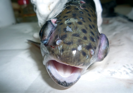
Fig. 1. Infectious exophthalmia and ocular hemorrhagic lesions in farmed rainbow trout (photo by T. A. Karaseva)
Exophthalmia and hemorrhages in the eyes of aquacultured rainbow trout were indicated by D. W. Bruno et al. in cases of acute viral hemorrhagic septicemia (VHS). VHS is widespread in Europe, North America, Japan, and Taiwan [10]. Similar pathology is observed in plasmocytoid leukemia of chinook salmon (Oncorhynchus tshawytscha) along the western coasts of America and Canada. The disease causative agent in salmonids is a retrovirus (salmon leukemia virus, SLV). Exophthalmia and subsequent blindness are typical for fish infected with viral encephalopathy and retinopathy (VER, or viral nervous necrosis, VNN) [14][16].
In 1980s, an outbreak of a new disease was reported in juvenile Caspian trout (Salmo trutta caspius) at the Chaikend fish hatchery, Azerbaijan SSR, when specific eye lesions were observed. Clinical examination demonstrated dense white papule-like structures (1–2 mm in diameter) with broad bases and tapered tips on the cornea and periorbital skin fold. As a result of their disintegration, the cornea and the periorbital skin fold degraded, which resulted in the discharge of the eye contents and eyeball prolapse. It was histologically determined that papule-like structures consist of epithelium, Bowman’s membrane and the corneal stroma. Eosinophilic and small basophilic inclusions were observed in the cytoplasm of the epithelial cells. Clusters of virus-like particles (30–40 nm) were detected using electron microscopy. The experts suggested these particles to be a virus of the Picornaviridae family [4][17].
In the early 1990s, fish pathologists A. M. Marchenko and T. E. Rodina described a coinfection in Caspian trout underyearlings and broodstock, which was caused by Renibacterium salmoninarum bacterium (causative agent of bacterial kidney disease) and an unknown filterable agent, presumably a virus. The diseased underyearlings demonstrated papule-like formations in the eyes similar to those detected in fish at the Chaikend fish hatchery, pale gills, sandy-colored liver, and gray edematous kidneys. The highest mortality rate (25%) was reported in fish with the specified clinical signs. Thorough disinfection of fish tanks and equipment, selection of clinically healthy broodstock, and feeding therapeutic feed with erythromycin helped to stop the fish mortality. However, for a long time, the ocular pathology was reported in singular individuals [4].
It is important that in the coming years, these diseases were not reported in the Caspian trout, and the suspected viral pathogens did not spread over the territory of the Russian Federation and were not reported in salmonids in other regions. Perhaps their spread was limited to the Caspian Sea basin. The viruses are known to lack epizootic potential in the ecosystem due to the rare contact of individuals of the same species, but with a high density of fish in aquaculture they can acquire pronounced pathogenic properties and trigger a disease outbreak [18].
Exophthalmia, hemorrhages in the eyeball and a wide range of signs of chronic pathology are reported among the symptoms of bacterial diseases [8][19-21]. For example, in 1986, at the Taibolsky fish hatchery in the Murmansk Oblast, a specific ocular pathology was first detected in juvenile Atlantic salmon (Salmo salar). Uncontrolled fish transportation resulted in rapid disease spread over the salmon hatcheries in the Murmansk and Arkhangelsk Oblasts and in Karelia [22][23]. In aquaculture practice, this pathology was reported as exophthalmia, nonparasitic cataract, or mechanical injury. The disease was observed in salmonids of all age groups raised in fish hatcheries: fry, underyearlings and two-year-olds [5][22][23]. The disease-typical signs were also detected in rainbow trout (Parasalmo mykiss) when grown in marine net cages, Eurasian river perch (Perca fluviatilis), common minnow (Phoxinus phoxinus) and ninespined stickleback (Pungitius pungitius), which inhabit freshwater lakes – water sources of the salmon hatcheries [22][23].
The bioassay results indicated that exophthalmia in Northwest fish hatcheries was caused by gram-positive cocci bacteria, which were initially identified as Streptococcus sp. In terms of biochemical properties, 93 of the obtained serotypes were homogeneous and in terms of antigenic properties they were close to the causative agent of streptococcal infection in yellowtail [22]. The disease was called streptococcal infection in salmonids, respectively. Later, in the 9th edition of “Bergey’s Manual of Determinative Bacteriology” (1993), these bacteria, pathogenic for salmonids and yellowtail, were assigned to the species Enterococcus seriolicida [24]. Thus, it was found that pathological processes in the eyes of fish developed in case of streptococcal infection. The disease cause is generally similar to septicemia throughout the year, so ocular pathology, which is only one of the enterococcal infection signs, develops gradually. At the beginning of the disease, the clinical signs are mainly manifested by unilateral exophthalmia and hemorrhages in the eyeball. Later, at different stages of the pathological process development, optic neuritis, corneal ulceration, vitreous prolapse and lens extrusion via pupillary rupture are reported, or leukoma is formed in fish. In the terminal disease stage, complete ocular prolapse occurs with conjunctival rupture [25]. Herewith, underyearlings salmonids do not survive, and in older fish the eye socket may become filled with pigmented connective tissue. Histological examination of the diseased fish revealed the fundus and iris hyperemia, corneal keratinization, delamination, erosion and necrosis, hyperemia and hemorrhages in the choroid, retinal deformity [22]. A number of authors find much in common in the epizootology of streptococcosis and such diseases as furunculosis and bacterial kidney disease, the etiological agents of which are closely associated with the hosts, and outside the body of fish can survive only for a limited time in water and bottom sediments [26].
At the same time, in the 1980s and 1990s, the juvenile salmonids demonstrated cataracts, the etiology of which could not be established. Thus, at the Petrozavodsky fish hatchery, singular cases of ocular lesions were reported in the juvenile salmon and lake salmon (Salmo salar morpha sebago); at the Vygsky fish hatchery, cataracts were observed in 8% of fish, and at Kemsky fish hatchery – in 9% of the total number of fish grown at the hatchery. No mortality was reported in the diseased fish. The microbiological test results did not confirm the infectious nature of cataracts in fish on Karelian aquaculture farms, though it was assumed against the background of widespread streptococcal infection [5].
Another bacterial disease that often affects fish’s eyes is vibriosis. It is a widespread disease of wild and aquacultured fish in marine and brackish waters [27-30]. Cold water vibriosis usually refers to Listonella (Vibrio) anguillarum-associated septicemia (Bergman, 1909). This Vibrio species represents aquatic saprotrophic microflora, it can be found in water, soil, mollusks and other marine inhabitants [9][31][32]. The main route of the infection transmission is with water and through contact with the diseased fish. On marine farms, Listonella anguillarum is released into the marine environment from the intestines, kidneys, ulcers and damaged eyes of the diseased and recovering fish. Serous hemorrhagic inflammation and tissue necrosis are typical at all stages of vibriosis. Of all the salmonids aquacultured in Europe, rainbow trout is the most susceptible to the disease. In the 1970s and 1980s, during the period of intensive development of trout aquaculture, vibriosis was widespread in the Gulf of Finland and Gulf of Riga located in the Baltic Sea. The disease outbreaks were reported in the farmed trout nearly every year, averaging a 30% mortality rate [33]. In northern European Russia, a vibriosis outbreak was first reported in two-year-old rainbow trout at White Sea fish farms in July 2004, two weeks after their transfer to marine cages. The disease was acute, and the mortality rate exceeded 40% [34].
Unilateral exophthalmia is generally observed in infected fish at the initial vibriosis stages. The subsequent ocular lesions are characterized by degeneration of all eye structures and tissues. They involve destruction of the cornea, dislocation of the lens, eyeball erosion and ulceration, and bleeding (Fig. 2). Less frequently, fish develop leukoma. In the survived individuals, the affected eye usually remains in place, with residual tissues staying within the eye socket [35].
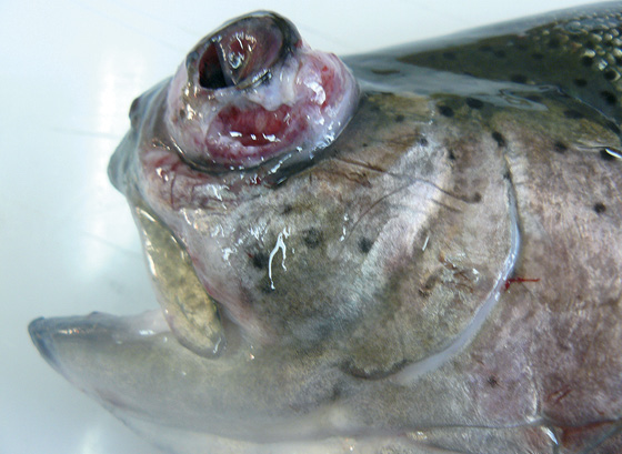
Fig. 2. Exophthalmia, corneal necrosis, and eyeball ulceration in rainbow trout with vibriosis caused by Listonella (Vibrio) anguillarum (photo by T. A. Karaseva)
Alimentary diseases. Since the early 1990s, the increase in the number of ocular pathologies in fish coincided with the introduction of granular salmon feeds and attempts to replace high-quality animal proteins in these feeds with plant proteins or low-quality animal protein substitutes. For the growth of fish, especially salmonids, use of balanced feed in the diet is very important. A deficiency of even one component in the feed leads to the development of changes, often irreversible, in the body of fish, including those in the eyes [36][37]. When using unbalanced feeds, the body is deficient in vitamins, amino acids, and mineral elements, which causes various types of eye lesions: cataracts, keratopathy, and eyeball protrusion. The S. G. Hughes review on ocular diseases that occur due to imperfect salmon feeds examines six types of pathologies. They include deficiency of riboflavin (vitamin B2), thiamine (vitamin B1), vitamin A, sulfur-containing amino acids (methionine and cysteine), tryptophan and zinc. In case of ocular pathologies, common are three main signs: cataracts, keratopathy, and exophthalmia. Furthermore, both eyes are affected [2].
Thus, in case of riboflavin (vitamin B2) deficiency in feed, lens opacity and sometimes corneal rupture with adhesion of the lens to cornea were reported in fish. Histological examination revealed corneal thickening, as well as vascularization, hyalinization and degeneration of the lens subepithelial layers. All these changes led to the loss of its transparency. The consequences of thiamine deficiency (vitamin B1) in salmonids involved corneal opacity and inflammation, blindness. Pathological changes in the eyes were noted in experimental conditions simulating lack of vitamin A in the diet of char and rainbow trout. Corneal and lens opacification as well as photophobia were reported in rainbow trout. The addition of beta-carotene to the feed prevented eye lesions only in warm water (above 12.4 °C), no such therapeutic effect was, however, observed at lower water temperatures [36]. Exophthalmia occurred with a lack of ascorbic acid and tocopherol (vitamin E) [37][38].
Ocular pathologies in fish are caused by a deficiency of sulfur-containing amino acids in the feed: methionine, cysteine, tryptophan. Their deficiency contributes to the lens opacification and involves the adjacent ocular tissues in the degenerative process. Japanese scientists found that when fed a zinc-deficient diet, the rainbow trout developed cataracts. The cataract that had already appeared did not resolve even when the fish began to receive feed with a sufficient amount of zinc. The zinc requirement for fish is 15–30 mg/kg of feed [36][39].
Corneal and lens opacification was reported when mold fungi were detected in the eyes of juvenile Atlantic salmon. The source of infection was substandard granular feed, and the disease was systemic. Moreover, the mold fungi formed mycelia in the fish eyes [40].
Since early 21st century, solving the problem of adequate feeding in the aquaculture of valuable fish species (salmon, whitefish) has significantly reduced the number of fish with ocular damage and improved the disease situation in salmon hatcheries in the Murmansk Oblast and Republic of Karelia.
Water toxicoses. The aquatic environment is often polluted by petroleum products, pesticides, chemical dyes, nitrates, and heavy metal salts [41]. In aquaculture, the fish lens is used as a test organ to assess the toxicity of chemical and other compounds [42, 43]. Experimental studies were conducted in this area, which demonstrated that the fish lens was highly sensitive to anthropogenic factors. The effects of such industrial toxicants as trichlorobenzene, nitrobenzene, 3-naphthol and salts of heavy metals (lead, copper, zinc) on fish were examined. It was found that these toxic compounds caused a change in proliferative activity in the lens epithelium [44]. The effect of toxicant exposure on cytodifferentiation, as well as on changes in the biochemical composition and optical properties in the lens nucleus and cortex (decreased optical density of the lens nucleus) was established. The peculiarities of the reaction of various cytodifferentiation zones of the fish lens epithelium to a number of toxic compounds (benzene, inorganic and heavy metal salts) are also reported, which consist in the inhibition or stimulation of mitotic activity in the germinal zone of the lens epithelium [41]. The formation and proliferation of tissue fibers further leads to cataracts.
Thus, the toxic effects of the aquatic environment also contribute to the development of pathological processes in the eyes of fish.
In the North-West of the Russian Federation, the Onezhsky fish hatchery was the most affected in terms of ocular pathologies for several years (2017–2022). According to the results of our long-term research, the main cause of the emerging pathologies in salmonids at this farm was the quality of the incoming water. Clinical investigation revealed exophthalmia in underyearling and two-year-old Atlantic salmon (Salmo salar) and brown trout (Salmo trutta). The highest level of eye lesions was reported in two-year-old Atlantic salmon – from 8 to 20%. In 2023, the percentage of fish with eye lesions was minimal (about 1%). The fish with unilateral or bilateral exophthalmia demonstrated gill pallor, pale and friable hepatic tissue, yellow bile in the gallbladder, and black discoloration of the posterior kidney. Microscopic examination of scrapings from the postorbital region of the protruded eyeball revealed a large number of coccoid and rod-shaped bacteria. After inoculation of the fundus contents on the nutrient media, the growth of colonies of bacteria of the genera Staphylococcus, Flavobacterium (Flexibacter), Pseudomonas was reported. The main source of water supply for the Onezhsky fish hatchery is lake Andozero. The meteorological conditions in summer lead to the lake shallowing, as a result of which the water entering the hatchery contains a large amount of inorganic suspensions. In summer, the water temperature rises and the oxygen level drops to 3.3–3.5 mg/L. It was perhaps these factors that contributed to the emergence and development of pathological processes in the eyes of salmonids with further exophthalmia, usually on the left side. To stabilize the water quality and temperature conditions in the pools, reconstruction of the water supply system with the installation of cooling equipment is essential.
When growing different types of fish in aquaculture conditions, there is a risk of various pathological processes. Among the factors contributing to the development of the pathological process in the eyes of fish, important are the following: pathogenic microorganisms, unbalanced feed, toxic substances. Despite the limited number of Russian and foreign publications on the problem of ocular pathology in salmonids, the literature review allows for the conclusion that the diseases leading to blindness in fish are an urgent problem for the fish hatcheries and farms all over the world.
In many descriptions of infectious and nutritional diseases, the term “exophthalmia” (eye protrusion) is used, so it is obvious that exophthalmia is a comprehensive term for many diseases and the most common pathological condition that can be observed in the eyes of fish.
In order to prevent any problems associated with ocular diseases, it is necessary to monitor the disease situation. An integrated approach is essential, which includes evaluation of the water source, monitoring of the incoming water quality during roe incubation and growing larvae to an adult, use of balanced feeds for salmonids, ichthyopathological monitoring of the immuno-physiological status of fish, and preventive measures to control infectious and parasitic diseases of fish. This will enable effective monitoring and minimize the risk of various ocular diseases in aquacultured salmonids.
1. Bruno D. W., Raynard R. S. The effect of water temperature on eye opacity in Atlantic salmon, Salmo salar L. Bulletin of the European Association of Fish Pathologists. 1994; 14 (3): 86–88.
2. Hughes S. G. Nutritional eye diseases in salmonids: a review. Progressive FishCulturist. 1985; 47 (2): 81–85. https://doi.org/10.1577/1548-8640(1985)47%3C81:NEDIS%3E2.0.CO;2
3. Ferguson H. Cataracts in fish – gross pathology and histopathology. Fish Pathology. 2022. https://fishhistopathology.com/?p=2697
4. Marchenko A. M., Rodina T. E. Smeshannaya infektsiya kaspiiskoi kumzhi, vyzvannaya Renibacterium salmoninarum i neizvestnym fil’truyushchim agentom = Mixed infection in Caspian trout caused by Renibacterium salmoninarum and an unidentified filterable agent. Parazity i bolezni ryb: tezisy dokladov IX Vsesoyuznogo soveshchaniya po parazitam i boleznyam ryb (Petrozavodsk, mart 1991 g.) = Parasites and diseases of fish: abstracts of the 9th AllUnion Conference on Fish Parasitology and Pathology (Petrozavodsk, March 1991). Leningrad; Zoological Institute Academy of Sciences of the USSR; 1990; 82–83. (in Russ.)
5. Mozharova A. I., Bychkova L. I. Sanitarno-epizooticheskoe sostoyanie lososevykh rybovodnykh zavodov Severnogo basseina = Sanitary and epizootic status of salmonid fish hatcheries in the Northern Basin. Rybnoe khozyaistvo. Seriya: Akvakul’tura: Bolezni ryb. Moscow: VNIERKH; 1996; (1): 1–7. (in Russ.)
6. Mirzoeva L. A. Epizooticheskoe sostoyanie lososevykh ferm Norvegii = Epizootic status of salmon farms in Norway. Rybnoe khozyaistvo. Seriya: Bolezni gidrobiontov v akvakul’ture. Moscow: VNIERKH; 2001; (1): 31–35. (in Russ.)
7. Hargis W. J. Disordes of the eye in finfish. Annual Review of Fish Diseases. 1991; 1: 95–117. https://doi.org/10.1016/0959-8030%2891%2990025-F
8. Shahin K., Veek T., Heckman T. I., Littman E., Mukkatira K., Adkison M., et al. Isolation and characterization of Lactococcus garvieae from rainbow trout, Onchorhyncus mykiss, from California, USA. Transboundary and Emerging Diseases. 2022; 69 (4): 2326–2343. https://doi.org/10.1111/tbed.14250
9. Egusa S. Infectious diseases of fish. New Delhi: Amerind Publishing Co. Pvt. Ltd.; 1992. 696 p.
10. Bruno D. W., Noguera P. A., Poppe T. T. A colour atlas of salmonid diseases. Springer Dordrecht; 2013. 211 p. https://doi.org/10.1007/978-94007-2010-7
11. Boerlage A. S., Elghafghuf A., Stryhn H., Sanchez J., Hammell K. L. Risk factors associated with time to first clinical case of Bacterial Kidney Disease (BKD) in farmed Atlantic salmon (Salmo salar L.) in New Brunswick, Canada. Preventive Veterinary Medicine. 2018; 149: 98–106. https://doi.org/10.1016/j.prevetmed.2017.11.014
12. Ferguson H. W., Hawkins L., MacPhee D. D., Bouchard D. Choroiditis and cataracts in Atlantic salmon (Salmo salar L) recovering from subzero water temperatures. Veterinary Record. 2004; 155 (11): 333–334. https://doi.org/10.1136/vr.155.11.333
13. Bjerkås E., Waagbø R., Sveier H., Breck O., Bjerkås I., Bjørnestad E., Maage A. Cataract development in Atlantic salmon (Salmo salar L) in fresh water. Acta Veterinaria Scandinavica. 1996; 37 (3): 351–360. https://doi.org/10.1186/bf03548101
14. Rahkonen R., Vennerström P., Rintamäki P., Kannel R. Terve kala, Tautien ennaltaehkäisy, tunnistus ja hoito. 2. uud. painos. Helsinki: Riista- ja kalatalouden tutkimuslaitos; 2013. 140 s. (in Finnish)
15. Mirzoyeva L. M. Virusnoe zabolevanie mozga i glaz molodi paltusa = Viral disease of brain and eyes in juvenile halibut. Rybnoe khozyaistvo. Seriya: Akvakul’tura: Bolezni ryb. Moscow: VNIERKH; 1997; (2): 24–26. (in Russ.)
16. Doan Q. K., Vandeputte M., Chatain B., Morin T., Allal F. Viral encephalopathy and retinopathy in aquaculture: a review. Journal of Fish Diseases. 2017; 40 (5): 717–742. https://doi.org/10.1111/jfd.12541
17. Marchenko A. M., Shchelkunov I. S., Kadoshnikov Yu. P. Obnaruzhenie virusopodobnogo agenta v porazheniyakh rogovitsy kaspiiskoi kumzhi = Detection of a virus-like agent in corneal lesions of Caspian trout. Nauchnotekhnicheskie problemy marikul’tury v strane: tezisy dokladov Vsesoyuznoi konferentsii (Vladivostok, 23–28 oktyabrya 1989 g.) = Scientific and technical challenges in mariculture: abstracts of the AllUnion Conference Proceedings (Vladivostok, 23–28 October 1989). Vladivostok: Pacific Research Institute for Fishery and Oceanography; 1989; 177–178. (in Russ.)
18. Alexyuk M., Alexyuk P., Bogoyavlenskiy A., Akanova K., Moldakhanov Y., Manakbayeva A., et al. Study of the diversity of fish viruses in the water area of the Central Caspian Sea by the method of metagenomic sequencing. Eurasian Journal of Ecology. 2023; 75 (2): 65–77. https://doi.org/10.26577/EJE.2023.v75.i2.06 (in Russ.)
19. Aguilar M., Isla A., Barrientos C. A., Flores-Martin S. N., Blanco J. A., Enríquez R., et al. Genomic and proteomic aspects of p57 protein from Renibacterium salmoninarum: Characteristics in virulence patterns. Microbial Pathogenesis. 2023; 174:105932. https://doi.org/10.1016/j.micpath.2022.105932
20. Kaur S., Kaur H., Kaur B., Naveen Kumar B. T., Tyagi A., Singh P., et al. Isolating pathogenic multidrug-resistant Aeromonas hydrophila from diseased fish and assessing the effectiveness of a novel lytic Aeromonas veronii bacteriophage (AVP3) for biocontrol. Microbial Pathogenesis. 2024; 196:106914. https://doi.org/10.1016/j.micpath.2024.106914
21. Irshath A. A., Rajan A. P., Vimal S., Prabhakaran V.-S., Ganesan R. Bacterial pathogenesis in various fish diseases: Recent advances and specific challenges in vaccine development. Vaccines. 2023; 11 (2):470. https://doi.org/10.3390/vaccines11020470
22. Karaseva T. A., Serdyuk A. V., Loginova G. A. Streptokokkovaya infektsiya na lososevykh khozyaistvakh Evropeiskogo Severa = Streptococcosis in salmonid aquaculture facilities of the European North. Sbornik nauchnykh trudov GosNIORKH. 1992; 331: 120–124. (in Russ.)
23. Karaseva T. A. Streptokokkoz lososevykh ryb = Streptococcal infection in salmonids. Rybnoe khozyaistvo. Seriya: Bolezni gidrobiontov v akvakul’ture. Moscow: VNIERKH; 2001; (1): 10–21. (in Russ.)
24. Holt J. G., Krieg N. R., Sneath P. H. A., Staley J. T., Williams S. T. Bergey’s manual of determinative bacteriology. 9th ed. Baltimore: Williams & Wilkins; 1994. 787 p.
25. Luo X., Fu X., Liao G., Chang O., Huang Z., Li N. Isolation, pathogenicity and characterization of a novel bacterial pathogen Streptococcus uberis from diseased mandarin fish Siniperca chuatsi. Microbial Pathogenesis. 2017; 107: 380–389. https://doi.org/10.1016/j.micpath.2017.03.049
26. Sindermann C. J. Disease in marine aquaculture. Helgoländer Meeresuntersuchungen. 1984; 37 (1–4): 505–532. https://doi.org/10.1007/BF01989327
27. Noga E. J. Fish disease: Diagnosis and treatment. 2nd ed. Wiley-Blackwell; 2010. 544 p. https://doi.org/10.1002/9781118786758
28. Austin B., Austin D. A. Bacterial fish pathogens. Diseases of farmed and wild fish. 4th ed. Chichester: Springer Praxis Publishing; 2007. 552 p. https://doi.org/10.1007/978-1-4020-6069-4
29. Egidius E. Vibriosis: pathogenicity and pathology. A review. Aquaculture. 1987; 67 (1–2): 15–28. https://doi.org/10.1016/0044-8486(87)90004-4
30. Kent M. L., Poppe T. T. Infectious diseases of coldwater fish in marine and brackish water. In: Diseases and Disorders of Finfish in Cage Culture. Ed. by P. T. K. Woo, W. Bruno, L. H. S. Lim. 2nd ed. Oxfordshire: CAB International; 2002; 61–105. https://doi.org/10.1079/9780851994437.0061
31. Lartseva L. V., Pivovarov Yu. P. Ecological epidemiology: a monograph. Astrakhan: Astrakhan State University; 2007. 187 p. (in Russ.)
32. Lartseva L. V. Prirodnaya ochagovost’ aeromonozov i vibriozov = Environmental persistence of aeromonosis and vibriosis. Ekologicheskie problemy prirodnykh i urbanizirovannykh territorii: materialy III Mezhdunarodnoi nauchnoprakticheskoi konferentsii (Astrakhan’, 20–21 maya 2010 g.) = Ecological challenges of natural and urbanized areas: proceedings of the 3rd International Science to Practice Conference (Astrakhan, May 20–21, 2010). Astrakhan: Astrakhan State University; 2010; 123–126. (in Russ.)
33. Vismanis К. О., Lullu F. V., Iygis V. A., Turovsky A. M., Yun A. I. Diseases of salmonids in Baltic Sea cages and their prevention. In: Biological foundations of aquaculture: fish parasites and diseases. Moscow: Nauka; 1984; 56–63. (in Russ.)
34. Karaseva T. A., Golikova L. N. Novye i redko vstrechayushchiesya bolezni raduzhnoi foreli (Parasalmo mykiss Walb.) = Emerging and rare diseases in rainbow trout (Parasalmo mykiss Walb.). Sostoyanie i puti razvitiya akvakul’tury v Rossiiskoi Federatsii: materialy IV Natsional’noi nauchnoprakticheskoi konferentsii (Kaliningrad, 8–10 oktyabrya 2019 g.) = State and development prospects of aquaculture in the Russian Federation: proceedings of the 4th National Science to Practice Conference (Kaliningrad, 8–10 October 2019). Saratov: Amirit; 2019; 117–121. https://elibrary.ru/bgarie (in Russ.)
35. Karaseva T. A. Patologii glaz u morskikh i presnovodnykh ryb Severnogo basseina = Ocular pathologies in marine and freshwater fish of the Northern Basin. Biological resources of the White Sea and inland waters of Euro pean North: Proceedings of the IV (XXVII) International conference (Vologda, December 5–10, 2005). Part 1. Vologda: Vologda State Pedagogical University; 2005; 170–172. (in Russ.)
36. Rodina T. E. Porazheniya glaz lososevykh ryb, obuslovlennye nedostatkom nekotorykh komponentov korma = Ocular lesions in salmonid fish associated with dietary component deficiencies. Rybnoe khozyaistvo. Seriya: Rybokhozyaistvennoe ispol’zovanie vnutrennikh vodoemov. Moscow: VNIERKH; 1989; (4): 6–13. (in Russ.)
37. Halver J. E., Smith R. R., Tolbert B. M., Baker E. M. Utilization of ascorbic acid in fish. Annals of the New York Academy of Sciences. 1975; 258 (1): 81–102. https://doi.org/10.1111/j.1749-6632.1975.tb29270.x
38. Wang K., Wang E., Qin Z., Zhou Z., Geng Y., Chen D. Effects of dietary vitamin E deficiency on systematic pathological changes and oxidative stress in fish. Oncotarget. 2016; 7 (51): 83869–83879. https://doi.org/10.18632/oncotarget.13729
39. Kubota S. S. Cataract in fishes: Pathological changes in the lens. Fish Pathology. 1976; 10 (2): 191–197. https://doi.org/10.3147/jsfp.10.191
40. Karaseva T. A., Golikova L. N. Otsenka zarazhennosti granulirovannykh kormov gribami i ikh rol’ v vozniknovenii patologii u kul’tiviruemykh lososevykh ryb = Assessment of fungal contamination in pelleted feeds and its role in pathology development in farmed salmonids. Current mycology in Russia: proceedings of the 4th Congress of Mycologists of Russia (Moscow, April 12–14, 2017). 2017; 7: 167–169. (in Russ.)
41. Lesnikov L. A. Razrabotka normativov dopustimogo soderzhaniya vrednykh veshchestv v vode rybokhozyaistvennykh vodoemov = Development of permissible limits for harmful substances in water of fishery reservoirs. Sbornik nauchnykh trudov GosNIORKH. 1979; 144: 3–41. (in Russ.)
42. Simakov Yu. G., Nikiforov-Nikishin A. L., Stebel’kov V. A., Arkhipov S. Yu. Izmeneniya soderzhaniya elementov v khrustalike danio i okunya pod vliyaniem zagryazneniya vodnoi sredy = Dynamics of elemental composition in zebrafish and perch lenses under aquatic pollution exposure. Vodnye bioresursy, vosproizvodstvo i ekologiya gidrobiontov: sbornik nauchnykh trudov = Aquatic biological resources, aquatic organism reproduction and ecology: collected scientific works. Moscow: VNIIPRKH; 1992; 66: 92–96. (in Russ.)
43. Nikiforov-Nikishin A. L., Kulaev S. N. Vozdeistvie toksikantov na dinamiku veshchestva u ryb = Toxicant exposure impacts on substance dynamics in fish. Vtoraya Vsesoyuznaya konferentsiya po rybokhozyaistvennoi toksikologii, posvyashchennaya 100letiyu problemy kachestva vody v Rossii (SanktPeterburg, noyabr’ 1991 g.): tezisy dokladov = Second AllUnion Conference on Aquaculture Toxicology devoted to 100year mark of water quality issue in Russia (Saint Petersburg, November 1991): proceedings. Saint Petersburg: GosNIORKH; 1991; 71–74. (in Russ.)
44. Nikiforov-Nikishin D. L. Morphological and biochemical aberrations in fish eye lenses induced by anthropogenic factors: Author’s abstract of thesis for degree of Cand. Sci. (Biology). Moscow; 2000. 23 p. (in Russ.)
Larisa I. Bychkova, Cand. Sci. (Biology), Senior Researcher, Department of Technology and Regulation of Aquaculture,
19, Okruzhnoy proezd, Moscow 105187.
Tatyana A. Karaseva, Cand. Sci. (Biology), Leading Researcher, Laboratory of Aquaculture and Diseases of Aquatic Organisms,
6, Akademika Knipovicha str., Murmansk 183038.
Vladimir A. Pylnov, Cand. Sci. (Biology), Leading Researcher, Department of Technology and Regulation of Aquaculture,
19, Okruzhnoy proezd, Moscow 105187.
Bychkova L.I., Karaseva, T.A., Pylnov V.A. Factors contributing to ocular pathologies in fish. Veterinary Science Today. 2025;14(2):140-147. https://doi.org/10.29326/2304-196X-2025-14-2-140-147
600901, Vladimir Oblast, Vladimir, microraion Yur’evets, ulitsa Gvardeyskaya, 6
FGBI “ARRIAH”
tel.: 8 (4922) 26-15-12, add. 22-27
E-mail: nikeshina@arriah.ru


