



Contents
Scroll to:
 V. A. Kalashnikova,
E. Yu. Radomskaya,
D. V. Bulgin,
V. I. Polyakova,
D. I. Dogadov,
A. V. Demerchyan,
N. V. Shcherbak,
D. V. Chukanov,
A. M. Goncharenko,
A. A. Minosyan,
I. M. Arshba
V. A. Kalashnikova,
E. Yu. Radomskaya,
D. V. Bulgin,
V. I. Polyakova,
D. I. Dogadov,
A. V. Demerchyan,
N. V. Shcherbak,
D. V. Chukanov,
A. M. Goncharenko,
A. A. Minosyan,
I. M. Arshba https://doi.org/10.29326/2304-196X-2026-15-1-28-37
Scroll to:
Introduction. Respiratory infections pose a significant challenge in veterinary practice due to their high prevalence across various animal species. Pneumonia and gastrointestinal diseases are leading causes of mortality in captive primates.
Objective. Study of pneumonia incidence in monkeys, the analysis of pulmonary microbiota composition, study of pathomorphological lesions in lung tissue.
Materials and methods. Common methods were used for pathomorphological, microscopic and bacteriological examinations of 1,862 dead monkeys. Lung samples and serum samples from 126 monkeys died of pneumonia in 2021–2024 were tested for acute respiratory viral pathogens as well as for antibodies to them with polymerase chain reaction (PCR) and enzyme-linked immunosorbent assay (ELISA).
Results. Pneumonia was postmortem diagnosed in 865 monkeys (46.5%). The mortality rate for pneumonia in baby monkeys during their first month of life reached 100%. In baby monkeys under one year of age, the mortality rate was 65.4%. The obtained data showed that the disease incidence in these age groups was high. Deaths of monkeys due to pneumonia were reported throughout the year. Based on postmortem examinations, bilateral polysegmental bronchopneumonia was the most frequent finding, lobar fibrinous pneumonia affecting the right lung was less common. Microscopic analysis detected purulent exudate and cocci bacteria in the bronchial lumen. The predominant bacteria isolated from lung tissue were enterobacteria (58.5%) and Gram-positive cocci (36.6%). Various microorganisms were isolated but the most frequently enteric bacteria were as follows: Escherichia coli (66.1%), Enterococcus spp. (27.5%) and Proteus spp. (31.5%). The following bacterial pathogens associated with pneumonia were detected: Staphylococcus aureus (31.5%), Klebsiella pneumoniae (2.2%), Pseudomonas aeruginosa (0.8%) and Streptococcus pneumoniae (0.6%). Adenoviruses, human parainfluenza viruses of type 1 and type 3 and respiratory syncytial virus (RSV) were also circulated in the monkey colony.
Conclusion. During analysis of microbial etiology of pneumonia in monkeys it shall be considered that pneumonia is frequently arisen as a secondary infection, heavily influenced by underlying gastrointestinal pathologies and immunosuppression.
Kalashnikova V.A., Radomskaya E.Yu., Bulgin D.V., Polyakova V.I., Dogadov D.I., Demerchyan A.V., Shcherbak N.V., Chukanov D.V., Goncharenko A.M., Minosyan A.A., Arshba I.M. Pathomorphological, bacteriological and virological features of pneumonia in captive monkeys. Veterinary Science Today. 2026;15(1):28-37. https://doi.org/10.29326/2304-196X-2026-15-1-28-37
In veterinary practice, pneumonia occurrence and treatment is of current importance due to the its high incidence rate among wild, domestic, farm animals (cats, dogs, cattle, pigs, horses, etc.) and birds. Pneumonia mainly affects baby and young animals, while the disease frequency and severity depend on the keeping conditions, stress, density of animals, their immune status, diet, climate and production practice, as well as coinfection with various pathogens [1][2][3][4][5][6][7][8][9][10][11]. In domestic animals, adults and elderly animals are often susceptible to the disease [4][5][6].
Low primates are also susceptible to most human pathogens, so it is easy to reproduce some human infectious diseases in them in a similar form [12]. In recent decades, due to the sharp decline in monkey populations in natural habitats and the ban on trapping, primate breeding centres have become the main source of laboratory primates. Animals kept in captivity are in most cases susceptible to intestinal and respiratory diseases. Pneumonia is one of the leading causes of morbidity and often leads to the death of animals [13][14][15]. Pneumonia may act either as an independent pathological process or as a concomitant (secondary) disease arising from gastrointestinal disorders, including acute or chronic gastritis and gastroenterocolitis [12][16]. In addition, pneumonia often occurs in animals recently imported from their natural habitats and undergoing acclimatization [17][18].
The lungs are a complex ecosystem with a large number of diverse microorganisms interacting both with each other and with the host organism [18][19]. A functional relationship exists between the microbiomes of the lungs, oropharynx, and intestines – an interconnection that significantly influences the development of both pulmonary and intestinal diseases through the modulation of metabolic, immune, and other physiological processes. Because of the ongoing microbial exchange between the oropharynx and the upper and lower respiratory tracts, the microbiome of the lungs is never static. The dominant microbial population in the microbiome and its size differ significantly between healthy and pathologically altered organs. According to this concept, infectious lung lesions are viewed as a disruption of the existing microbial balance, with the course and outcome of pneumonia being largely determined by interactions among the microorganisms themselves [19].
A critical step in pneumonia diagnosis is the isolation and identification of the involved infectious agents – including bacteria, viruses, and fungi – from the respiratory tract [2][20]. According to literature data, less than 10% of pneumonia cases are described as polymicrobial, and in more than 50% of cases, the etiological agents of pneumonia in monkeys remain unidentified [19]. According to the literature data, the most common causes of pneumonia in captive monkeys of various species are Streptococcus spp., less commonly Klebsiella pneumoniae, Staphylococcus aureus and other bacteria (Pseudomonas aeruginosa, Escherichia coli, Enterococcus spp., Morganella morganii) [21][22][23][24][25]. Earlier, staff-members of the Kurchatov Complex of Medical Primatology of National Research Centre “Kurchatov Institute” demonstrated the involvement of human parainfluenza virus type 3 in the respiratory tract pathology in Papio anubis [26].
The importance of this study is highlighted by the fact that respiratory diseases are predominant in the etiological structure of diseases in monkeys and constitute one of the primary causes of mortality for animals in captivity.
This study presents novel data by consolidating the results of pneumonia mortality monitoring in captive monkeys in the primate breeding centre, representing the first such investigation carried out in Russia. Accordingly, the present study was aimed at not only bacterial microflora detection in the lungs of dead animals and assessment of serological and molecular-genetic markers of acute viral respiratory infections (AVRIs), but also at description of pathologically altered lung features.
The purpose of the study was to study the pneumonia incidence, to analyse lung microbiota landscape, and to examine lung lesions.
Animals. A total of 1,862 deaths of monkeys of various species from spontaneous diseases were studied in the period from January 2019 to December 2024. All animals were kept in the primate breeding centre of the Kurchatov Complex of Medical Primatology of National Research Centre “Kurchatov Institute”. Dead male (n = 726) and female (n = 1,136) monkeys ranged in age from new-born to 38 years (Table 1).
Table 1
Characterization of dead monkeys by age groups
|
Monkey species |
Age groups |
Total |
|||||
|
under 1 month of age |
under 11 months of age |
1–3 years of age |
4–10 years of age |
11–15 years of age |
16 years of age and older |
||
|
Macaca mulatta |
52 |
45 |
116 |
176 |
86 |
129 |
604 |
|
Macaca fascicularis |
73 |
32 |
78 |
148 |
76 |
94 |
501 |
|
Macaca nemestrina |
15 |
5 |
2 |
8 |
6 |
17 |
53 |
|
Chlorocebus aethiops ssp. |
11 |
7 |
7 |
18 |
14 |
18 |
75 |
|
Papio anubis |
30 |
13 |
27 |
40 |
21 |
20 |
151 |
|
Papio hamadryas |
109 |
49 |
47 |
110 |
58 |
71 |
444 |
|
Other species |
5 |
2 |
7 |
9 |
3 |
8 |
34 |
|
Total |
295 |
153 |
284 |
509 |
264 |
357 |
1,862 |
Postmortem and microscopic examinations. Dead animals were necropsied in the necropsy room of the Laboratory for Pathological Anatomy of the Kurchatov Complex of Medical Primatology. Internal organs and tissues from dead animals were examined to detect pathological lesions. When gross lung inflammation signs were detected, tissue samples were collected for histological analysis. The samples were fixed in a 10% neutral (pH 7.4) formalin solution, then subjected to standard histological processing, followed by mounting into Histomix paraffin medium (BioVitrum, Russia). Histological sections, 4 μm thick, were prepared from the paraffin-embedded tissues and stained with Hansen’s hematoxylin and eosin, as well as with Van Gieson’s picrofuchsin [27].
Morphological analysis (microscopic examination) was carried out with Axiolab.A1 laboratory microscope (Carl Zeiss Microscope GmbH, Germany). Axiocam 105 colour digital camera (Carl Zeiss Microscopy GmbH, Germany) was used for microphotography.
Bacteriological examination was carried out according to a common technique: a lung smear was made on a slide, Gram-stained, and inoculated in diagnostic nutrient media, followed by biochemical identification of the grown colonies of microorganisms, as described earlier [28].
Virological examination. Serum and lung samples collected in 2021–2024 from 126 dead monkeys with diagnosed pneumonia including 48 Macaca mulatta, 22 Macaca fascicularis, 7 Chlorocebus aethiops ssp., 38 Papio hamadryas and 11 Papio anubis were used for the virological examination. The lung suspension was prepared using Minilys homogenizer (Bertin Technologies, France) at a ratio of 5–6 g of the material per 1 mL of 0.1M sodium phosphate buffer, pH 7.4; centrifuged with Allegra cold centrifuge (Beckman Coulter, USA) at 3,000 rpm for 30 min for clarification. The resulting 10% supernatant was used for further tests.
Test kits manufactured by ECOlab (Russia) were used for detection of IgG, IgM and IgA antibodies to parainfluenza virus types 1 and 3, respiratory syncytial virus (RSV) and adenovirus. The results of enzyme-linked immunosorbent assay (ELISA) were read using Immunochem-2100 laboratory spectrophotometer (High Technology Inc., USA) at wavelength of 450 nm. The sera reactivity to respiratory viruses was assessed based on OD450 values (optical density of ELISA-tested serum samples at a wavelength of 450 nm). The test results were interpreted according to the test-kit manufacturer’s instructions.
Nucleic acids were extracted from the prepared 10% lung supernatant using RIBO-prep kit (Central Research Institute of Epidemiology of Rospotrebnadzor, Russia) according to the manufacturer’s instructions. Complementary DNA was synthesized on a total RNA matrix using Reverta-L reagent kit (Central Research Institute of Epidemiology of Rospotrebnadzor, Russia) in accordance with the manufacturer’s instructions. Resulting cDNAs were amplified using “AmpliSens® AVRI-screen-FL” real-time PCR kit to identify acute respiratory viral infection pathogens (RSV; metapneumovirus; parainfluenza virus types 1, 2, 3 and 4; coronavirus; rhinovirus; adenovirus groups B, C and E; bocavirus) according to the manufacturer’s instructions. Amplification and analysis of the results were performed using Rotor-Gene Q device (QIAGEN GmbH, Germany).
Statistical data processing. Statistical processing of the data and calculations were carried out using GraphPadPrism 8 software. To detect changes in frequency metrics across study years or age groups, a χ² trend test (Pearson’s chi-square) was applied. All differences were interpreted as significant at p < 0.05.
A total of 865 pneumonia cases (46.5% of the total number of dead animals) were detected over a six-year period based on the results of postmortem examinations (Table 2). Pneumonia was more frequently diagnosed in males than in females (52.8 and 42.4%, respectively). Pneumonia mortality rates were generally similar across monkey species, exception for Papio hamadryas and Macaca nemestrina. In these monkey species, pneumonia caused more than 50% of deaths.
Table 2
Characterization of fatal pneumonia cases in monkeys
|
Monkey species |
Quantity/% |
||
|
total |
females |
males |
|
|
Macaca mulatta |
249/41.2 |
156/40.7 |
93/42.1 |
|
Macaca fascicularis |
221/44.1 |
129/39.5 |
92/52.9 |
|
Macaca nemestrina |
33/62.3 |
21/70.0 |
12/52.2 |
|
Chlorocebus aethiops ssp. |
32/42.7 |
22/44.0 |
10/40.0 |
|
Papio anubis |
74/49.0 |
37/40.2 |
37/62.7 |
|
Papio hamadryas |
236/53.2 |
109/45.8 |
127/61.7 |
|
Other species |
20/58.8 |
8/50.0 |
12/66.7 |
|
Total |
865/46.5 |
482/42.4 |
383/52.8 |
According to the findings, the highest mortality for pneumonia was observed in monkeys during their first month of life (Table 3).
Table 3
Characterization of monkeys died of pneumonia by age groups
|
Monkey species |
Age groups |
p, χ² for trend* |
|||||||||||
|
under 1 month of age |
under 11 month of age |
1–3 years of age |
4–10 years of age |
11–15 years of age |
16 years of age and older |
||||||||
|
number of animals |
% |
number of animals |
% |
number of animals |
% |
number of animals |
% |
number of animals |
% |
number of animals |
% |
||
|
Macaca mulatta |
47 |
90.4 |
25 |
55.6 |
46 |
39.7 |
62 |
35.2 |
24 |
27.9 |
45 |
34.9 |
0.0005↑↓ |
|
Macaca fascicularis |
72 |
98.6 |
22 |
68.8 |
22 |
28.2 |
41 |
27.7 |
23 |
30.3 |
41 |
43.6 |
0.7675 |
|
Macaca nemestrina |
14 |
93.3 |
1 |
20.0 |
1 |
50.0 |
6 |
75.0 |
4 |
66.7 |
7 |
41.2 |
0.9643 |
|
Chlorocebus aethiops ssp. |
9 |
81.8 |
1 |
14.3 |
1 |
14.3 |
5 |
27.8 |
6 |
42.9 |
10 |
55.6 |
0.0258↓↑ |
|
Papio anubis |
27 |
90.0 |
9 |
69.2 |
9 |
33.3 |
13 |
32.5 |
8 |
38.1 |
8 |
40.0 |
0.1357 |
|
Papio hamadryas |
100 |
91.7 |
40 |
81.6 |
29 |
61.7 |
25 |
22.7 |
17 |
29.3 |
25 |
35.2 |
< 0.0001↓ |
|
Other species |
5 |
100 |
2 |
100 |
5 |
71.4 |
4 |
44.4 |
1 |
33.3 |
3 |
37.5 |
0.9648 |
|
Total |
274 |
92.9 |
100 |
65.4 |
113 |
39.8 |
156 |
30.6 |
83 |
31.4 |
139 |
38.9 |
< 0.0001↑↓ |
* p < 0.05 (χ² test – statistical difference in pneumonia diagnosis rates between monkey species). Arrows indicate the trend in detection frequency by age, providing that the test result was statistically significant.
The total number of monkeys that died due to pneumonia averaged between 126 and 166 cases per year, remaining relatively stable in percentage terms over a six-year period (Table 4).
Table 4
Number of monkeys died of pneumonia by year
|
Monkey species |
Year of testing |
p, χ² for trend* |
|||||
|
2019 |
2020 |
2021 |
2022 |
2023 |
2024 |
||
|
Macaca mulatta |
42 |
32 |
43 |
36 |
44 |
52 |
0.0075↑↓ |
|
Macaca fascicularis |
42 |
44 |
39 |
27 |
32 |
37 |
0.4535 |
|
Macaca nemestrina |
6 |
9 |
3 |
3 |
8 |
4 |
0.7173 |
|
Chlorocebus aethiops ssp. |
5 |
5 |
2 |
6 |
10 |
4 |
0.3061 |
|
Papio anubis |
20 |
8 |
15 |
7 |
14 |
10 |
0.3494 |
|
Papio hamadryas |
43 |
42 |
40 |
45 |
38 |
28 |
0.3828 |
|
Other species |
8 |
4 |
3 |
2 |
1 |
2 |
0.0177↓ |
|
Total |
166/46.6% |
144/45.4% |
145/38.2% |
126/49.2% |
147/51.9% |
137/50.8% |
0.0910 |
* p < 0.05 (χ² test – statistical difference in pneumonia diagnosis rates between monkey species). Arrows indicate the trend in detection frequency by year, providing that the test result was statistically significant.
Over the six-year study period, neonatal mortality in monkeys exhibited a steady increase, with pneumonia accounting for 100% of deaths in this age group by 2024.
Analysis of seasonal patterns in monkey mortality due to pneumonia revealed that more than 50% of cases occurred during the autumn-winter period of 2020 and 2024, and during the summer-spring period of 2023. In 2022, mortality due to pneumonia in these animals remained high throughout all seasons.
Pathomorphological features of pneumonia. Polysegmental bronchopneumonia was more frequently observed in monkeys living at the primate breeding centre, whereas lobar fibrinous pneumonia – similar to human croupous pneumonia – was considerably less common. Macroscopically, bilateral pulmonary involvement was the most common finding, with the right lung being affected more extensively or more frequently. In cases of unilateral involvement, pneumonia was observed more frequently in the right lung alone (Fig. 1).
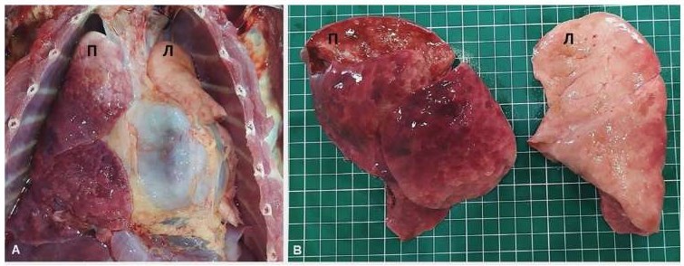
Fig. 1. Macrophotography of heart – lung complex in the monkey with polysegmental bronchopneumonia (Macaca fascicularis, ♀, 3 years old): A – general view; B – all lobes of the right lung are affected; П – right lung; Л – left lung
Croupous pneumonia in monkeys was characterized by lobar fibrinous inflammation with pleura lesions. In addition to lobar involvement, large focal pneumonic lesions were detected in the centre of the lobes or extending to the pleura. The affected lung tissue was airless, of dense consistency and greyish-reddish colour. A cloudy, foamy fluid exuded from the cut surface when the affected lung was compressed. The lung lymph nodes were enlarged. Cases of total, bilateral lung damage with alveolar filling by fibrinous-leukocytic exudate and serous fluid buildup were recorded (Fig. 2).
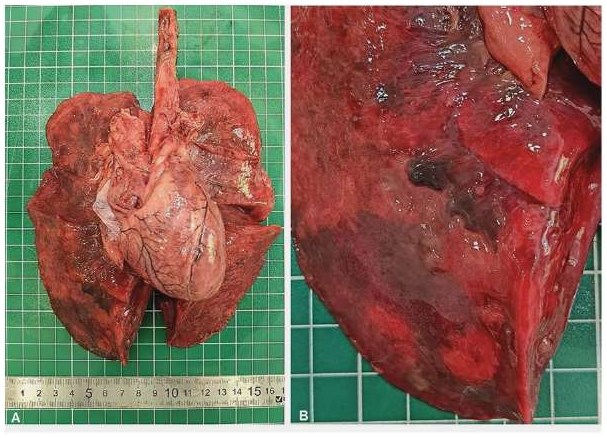
Fig. 2. Macrophotography of heart – lung complex in the monkey with fibrinous pneumonia (Erythrocebus patas, ♂, 10 years old), all lobes of the right and left lungs are completely affected: A – general view; B – fibrin threads on the visceral pleura of the right lung
Fibrin, degenerated leukocytes, and other cell debris were detected within the alveoli in croupous pneumonia cases. There were areas of lung tissue containing only red blood cells in the alveoli. Apparent vascular hyperemia with parietal leukocyte accumulation and capillary stasis within the interalveolar septa were observed (Fig. 3).

Fig. 3. Microscopic lesions in lungs of the monkey with lobar pneumonia (Macaca mulatta, ♂, 5 years old): A – alveolar edema, fibrin threads in the alveolar lumen, degenerated leukocytes (hematoxylin and eosin staining, magnification 200×); B – alveolar edema, erythrocyte and leukocyte accumulations in alveolar lumen (hematoxylin and eosin staining, magnification 400×)
Purulent tissue melting foci located generally around the affected bronchi were observed in all croupous pneumonia cases. The bronchial lumen contained purulent exudate and coccoid microflora.
Bronchopneumonia occurred both independently and as a concomitant disease complicating gastrointestinal pathologies. In bronchopneumonia cases, multiple small inflammation foci of grey-red or bluish-purple colour, often merging, that located along the branching bronchi were found in the lungs. The lung tissue around inflammation foci was edematous with hyperemia or severe emphysema, which gave the incision surface a mottled appearance (Fig. 4).
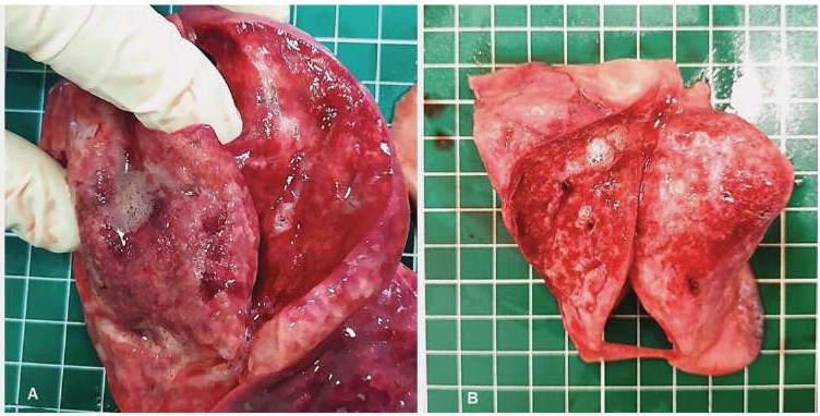
Fig. 4. Macroscopic lesions in lungs of the monkey with bronchopneumonia (Papio anubis, ♂, 5 years old): A, B – alveolar edema (foamy fluid on section), mottled pattern in lung tissue
Similar to croupous pneumonia, the microscopic inflammation lesions in these cases were characterized by accumulation of the exudate of various types. Foci of serous fluid mixed with red blood cells as well as polymorphic cellular exudate were found in the alveoli. Leukocytes and mucous with a large amount of bacterial microflora often predominated in the exudate. In all cases, inflammatory lung lesions were combined with the development of focal atelectasis and focal emphysema (Fig. 5).
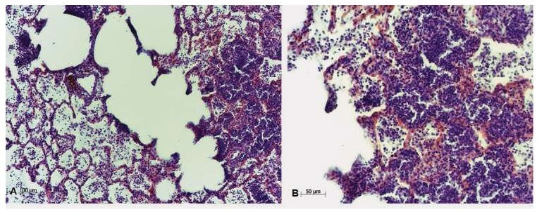
Fig. 5. Microscopic lesions in lungs of the monkey with bronchopneumonia (Papio anubis, ♂, 5 years old): A – focal emphysema (dilated alveoli, thinned and partly disrupted alveolar walls), polymorphic cellular exudate in the alveoli lumen (hematoxylin and eosin staining, magnification 100×); B – polymorphic cellular exudate in the alveoli lumen (hematoxylin and eosin staining, magnification 200×)
Thus, croupous pneumonia and bronchopneumonia in monkeys kept in the Kurchatov Complex of Medical Primatology primate breeding centre were characterized by a variety of morphological changes in the lungs. This appears related to the different properties of the disease pathogens.
Asymptomatic pneumonia diagnosed at necropsy only were found in most dead animals. In some cases, differentiation between croupous pneumonia and bronchopneumonia was difficult, as the presenting symptoms were subtle or nonspecific. Intrapulmonary complications including purulent bronchitis, lymphangitis, purulent inflammation foci, extremely rarely – pleural empyema were observed in some animals. The following extrapulmonary lesions were recorded – purulent meningitis, serous-purulent pericarditis.
Microbial landscape in lungs. Gram-positive cocci were found in all smears during examination of lung microbiota of monkeys. Over six years, 1,660 microorganisms were isolated during bacteriological tests, the majority of which were representatives of the family Enterobacteriaceae (58.5%). Gram-positive cocci were detected in 36.6% of cases, the proportion of non-fermenting bacteria, including Pseudomonas aeruginosa, and non-differentiated Gram-positive rods was 1.0 and 3.8%, respectively (Table 5).
Table 5
Number of microorganisms isolated from lungs of the monkeys died of pneumonia (2019–2024)
|
Microorganism |
Year of testing |
p, χ² for trend |
Total |
|||||
|
2019 |
2020 |
2021 |
2022 |
2023 |
2024 |
|||
|
E. coli |
112 |
100 |
106 |
85 |
87 |
82 |
0.2274 |
572 |
|
Representatives of tribe Proteeae |
51 |
50 |
52 |
42 |
62 |
44 |
0.0002↑↓ |
301 |
|
Klebsiella spp. |
18 |
8 |
5 |
4 |
3 |
3 |
0.0048↓ |
41 |
|
Enterobacter spp. |
7 |
4 |
4 |
1 |
4 |
6 |
0.5613 |
26 |
|
Citrobacter spp. |
7 |
4 |
0 |
2 |
2 |
0 |
0.0344↑↓ |
15 |
|
Other enterobacteria |
4 |
5 |
1 |
4 |
4 |
1 |
0.9126 |
19 |
|
Ps. aeruginosa |
1 |
2 |
2 |
0 |
1 |
1 |
0.9926 |
7 |
|
Other non-fermenting bacteria |
0 |
0 |
1 |
0 |
2 |
6 |
< 0.0001↑ |
9 |
|
Bacillus spp. |
1 |
1 |
6 |
4 |
3 |
7 |
0.0024↓↑ |
22 |
|
Other Gram-positive rods |
27 |
10 |
0 |
0 |
3 |
2 |
< 0.0001↓ |
42 |
|
Staphylococcus spp. |
123 |
64 |
41 |
40 |
62 |
25 |
< 0.0001↓ |
355 |
|
Enterococcus spp. |
53 |
49 |
40 |
31 |
36 |
29 |
0.9856 |
238 |
|
Other Gram-positive cocci, including Streptococcus pneumoniae |
6 |
3 |
0 |
1 |
2 |
0 |
0.0503 |
12 |
|
Candida spp. |
0 |
0 |
1 |
0 |
0 |
0 |
0.9320 |
1 |
|
Total |
410 |
300 |
259 |
214 |
271 |
206 |
< 0.0001↓ |
1,660 |
* p < 0.05 (χ² test – statistical difference in detection frequency between microorganism species). Arrows indicate the trend in detection frequency by year, where the test result was statistically significant.
Statistical analysis showed significant changes in the detection rates of certain microorganisms over the years of the study (p < 0.0001), which indicated shifts in lung microbiota composition.
The number of representatives of tribe Proteeae varied without any clear tendency to increase or decrease, which was indicative of a dynamic change in the role of these microorganisms (p = 0.0002). The number of Klebsiella spp. detections decreased over the years (p = 0.0048), potentially indicating a decline in the pathogen’s role in pneumonia development. The number of Citrobacter spp. (p = 0.0344) and Bacillus spp. (p = 0.0024) detections varied over the years. Notwithstanding the high isolation rate of Staphylococcus spp., a significant decline has been recorded in recent years, from 123 to 25 cases (p < 0.0001). Gram-positive rods detection rate also decreased (p < 0.0001). Non-fermenting Gram-negative rods were rarely isolated from the lungs, however, they were detected in 6 cases (p < 0.0001) in 2024. The total level of the isolated microorganisms decreased over the years (from 410 in 2019 to 206 in 2024), which was confirmed by statistical data (p < 0.0001).
As for microbial landscape, the leading position was occupied by E. coli, which were found in the lungs of 66.1% of monkeys, the second position was occupied by Staphylococcus spp., including S. aureus (41.1%), followed by representatives of tribe Proteeae (Proteus spp., Providencia spp., M. morganii) and Enterococcus spp. (34.8 and 27.5%, respectively). Other microorganisms were isolated rarely. During the tested period, S. aureus was more often isolated as compared to other main bacterial pathogens of pneumonia – in 267 monkeys (31.5%), other pathogens were found in some cases, namely: K. pneumoniae – in 19 monkeys (2.2%), Ps. aeruginosa – in 7 monkeys (0.8%), St. pneumoniae – in 5 monkeys (0.6%). No bacterial growth was observed on the nutrient media inoculated by the samples from 7 cases (0.8%). Also, no materials were collected for examination from 17 monkeys with pneumonia due to postmortem decomposition. Bacterial associations were observed, however, the number of detected associations decreased during the tested period. In 2019, 89% of the detected microbial isolates were associations, compared to 52% in 2024, with the frequency of 4-component associations apparently decreasing.
Any analysis of the etiological role of the isolated microorganisms in monkey pneumonia shall account for the fact that, in the majority of cases, the disease has arisen concurrently with gastrointestinal pathology and immunosuppression. This was evidenced by the detection of coliforms in lungs, as well as the prolonged pneumonia course with subtle or non-specific clinical signs. S. aureus represented an exception, based on previous molecular genetic evidence indicating its high pathogenic potential and lung tissue tropism of some strains [29][30].
When 126 monkeys with diagnosed pneumonia who died in 2021–2024 were tested for AVRI indicators, IgG antibodies indicative of post-infection immunity against the following viruses were detected: parainfluenza virus type 1 and 3 (14.3 and 4.0% of the monkeys, respectively), RSV (1.2%), and adenovirus (8.7%). IgA and IgM antibodies indicative of acute infection period were detected only against RSV (35.3% of the monkeys). No AVRI pathogen RNAs/DNAs were detected with polymerase chain reaction (PCR) in the lung parenchyma (Table 6).
Table 6
Indicators of viral infections
|
Indicators Monkey species |
Parainfluenza virus type 1 |
Parainfluenza virus type 3 |
RSV |
Adenovirus |
|||
|
IgG |
IgA |
IgG |
IgA |
IgG |
IgM |
IgG |
|
|
Monkeys that died in 2021–2022 |
|||||||
|
Macaca mulatta |
10/15* 66.7% |
0/15 0% |
0/15 0% |
0/15 0% |
n/d |
n/d |
2/15 13.3% |
|
Macaca fascicularis |
3/4 75.0% |
0/4 0% |
0/4 0% |
0/4 0% |
n/d |
n/d |
0/4 0% |
|
Chlorocebus aethiops ssp. |
1/3 33.3% |
0/3 0% |
0/3 0% |
0/3 0% |
n/d |
n/d |
0/3 0% |
|
Papio anubis |
3/15 20.0% |
0/15 0% |
0/15 0% |
0/15 0% |
n/d |
n/d |
0/15 0% |
|
Papio hamadryas |
0/4 0% |
0/4 0% |
0/4 0% |
0/4 0% |
n/d |
n/d |
0/4 0% |
|
Total |
17/41 41.5% |
0/41 0% |
0/41 0% |
0/41 0% |
n/d |
n/d |
2/41 4.9% |
|
Monkeys that died in 2023–2024 |
|||||||
|
Macaca mulatta |
0/33 0% |
0/33 0% |
0/33 0% |
0/33 0% |
0/33 0% |
8/33 24.2% |
4/33 12.1% |
|
Macaca fascicularis |
1/18 5.6% |
0/18 0% |
0/18 0% |
0/18 0% |
0/18 0% |
9/18 50.0% |
1/18 5.6% |
|
Chlorocebus aethiops ssp. |
0/4 0% |
0/4 0% |
2/4 50.0% |
0/4 0% |
1/4 25.0% |
2/4 50.0% |
1/4 25.0% |
|
Papio anubis |
0/23 0% |
0/23 0% |
3/23 13.0% |
0/23 0% |
0/23 0% |
8/23 34.8% |
2/23 8.7% |
|
Papio hamadryas |
0/7 0% |
0/7 0% |
0/7 0% |
0/7 0% |
0/7 0% |
3/7 42.9% |
1/7 14.3% |
|
Subtotal |
1/85 1.2% |
0/85 0% |
5/85 5.9% |
0/85 0% |
1/85 1.2% |
30/85 35.3% |
9/85 10.6% |
|
Total |
18/126 14.3% |
0/126 0% |
5/126 4.0% |
0/126 0% |
1/85 1.2% |
30/85 35.3% |
11/126 8.7% |
* positive serum samples / number of tested serum samples; n/d – no data.
In monkeys that died in 2021–2022 (n = 41), IgG antibodies against parainfluenza virus type 1 (41.5%) only and adenovirus (4.9%) were detected, while no IgG antibodies against parainfluenza virus type 3 were detected. Also, no serological indicators of acute infection were detected.
In animals with diagnosed pneumonia that died in 2023–2024 (n = 85), IgM antibodies against RSV (35.3%) were detected in their sera, while the no IgA antibodies against parainfluenza virus type 1 and 3 were detected. In addition, IgG antibodies indicative of postinfection immunity were detected: IgG against parainfluenza virus type 1 and 3 (1.2 and 5.9%, respectively), RSV (1.2%), and adenovirus (10.6%).
1. Kostyleva O. A. Staphylococcosis in dogs and cats (clinical aspects, treatment). Bulletin of Altai State Agricultural University. 2005; (2): 49–52. https://elibrary.ru/ircoun (in Russ.)
2. Kozlova S. V., Krasnolobova E. P., Veremeeva S. A. On the question of fungal-bacterial associations of the respiratory system organs of birds. Vestnik of Kursk State Agricultural Academy. 2022; (8): 145–150. https://elibrary.ru/nizvrx (in Russ.)
3. Kudriashov A. A., Balabanova V. I., Pudovkin D. N., Belyaeva E. V. Pathologic diagnosis of infectious resppiratory diseases of cattle on farms. Actual Questions of Veterinary Biology. 2017; (1): 59–65. https://elibrary.ru/ygroux (in Russ.)
4. Shulga N. N., Shulga I. S., Dikunina S. S., Plavshak L. P. The spread of calves’ respiratory diseases in the Amur Region. Far Eastern Agricultural Journal. 2016; (3): 90–93. https://elibrary.ru/wxotyh (in Russ.)
5. Polishchuk S. V., Belyavtseva E. A. Diagnosis enzootic pneumonia pigs at farm “Veles-Crimea”. Transactions of Taurida Agricultural Science. 2015; (1): 164–171. https://elibrary.ru/wfdujf (in Russ.)
6. Naumov M. M., Kononova T. A. Infectious pneumonia in cats, analysis of incidence on the basis of the Kursk veterinary center “Beethoven”. Vestnik of Kursk State Agricultural Academy. 2022; (4): 82–85. https://elibrary.ru/bddknd (in Russ.)
7. Stephenson T., Lee K., Griffith J. E., McLelland D. J., Wilkes A., Bird P. S., et al. Pulmonary actinomycosis in South Australian koalas (Phascolarctos cinereus). Veterinary Pathology. 2021; 58 (2): 416–422. https://doi.org/10.1177/0300985820973459
8. Black S. R., Barker I. K., Mehren K. G., Crawshaw G. J., Rosendal S., Ruhnke L., et al. An epizootic of Mycoplasma ovipneumoniae infection in captive Dall’s sheep (Ovis dalli dalli). Journal of Wildlife Diseases. 1988; 24 (4): 627–635. https://doi.org/10.7589/0090-3558-24.4.627
9. Zhao W., Tian Q., Luo Y., Wang Y., Yang Z.-X., Yao X.-P., et al. Isolation, identification, and genome analysis of lung pathogenic Klebsiella pneumoniae (LPKP) in forest musk deer. Journal of Zoo and Wildlife Medicine. 2017; 48 (4): 1039–1048. https://doi.org/10.1638/2016-0241.1
10. McClure S., Sibert G., Hallberg J., Bade D. Efficacy of a 2-dose regimen of a sustained release ceftiofur suspension in horses with Streptococcus equi subsp. zooepidemicus bronchopneumonia. Journal of Veterinary Pharmaco logy and Therapeutics. 2011; 34 (5): 442–447. https://doi.org/10.1111/j.1365-2885.2011.01267.x
11. Schmidt V., Marschang R. E., Abbas M. D., Ball I., Szabo I., Helmuth R., et al. Detection of pathogens in Boidae and Pythonidae with and without respiratory disease. Veterinary Record. 2013; 172 (9):236. https://doi.org/10.1136/vr.100972
12. Krylova R. I. Sravnitel’naya kharakteristika nozologicheskogo profilya lyudei i obez’yan raznykh vidov = Comparative characterization of nosological profiles: humans vs monkeys of different species. Laboratornye primaty dlya resheniya aktual’nykh problem meditsiny i biologii: materialy simpoziuma = Laboratory Primates: Key Models for Modern Medical and Biological Research: Symposium Proceedings. Moscow: RAMS Publishing House; 2004; 18–21 (in Russ.)
13. Dick E. J. Jr., Owston M. A., David J. M., Sharp R. M., Rouse S., Hubbard G. B. Mortality in captive baboons (Papio spp.): a-23-year study. Journal of Medical Primatology. 2014; 43 (3): 169–196. https://doi.org/10.1111/jmp.12101
14. Bommineni Y. R., Dick E. J. Jr., Malapati A. R., Owston M. A., Hubbard G. B. Natural pathology of the baboon (Papio spp.). Journal of Medical Primatology. 2011; 40 (2): 142–155. https://doi.org/10.1111/j.1600-0684.2010.00463.x
15. Laurence H., Kumar S., Owston M. A., Lanford R. E., Hubbard G. B., Dick E. J. Jr. Natural mortality and cause of death analysis of the captive chimpanzee (Pan troglodytes): A 35-year review. Journal of Medical Primato logy. 2017; 46 (3): 106–115. https://doi.org/10.1111/jmp.12267
16. Lapin B. A., Dzhikidze E. K., Krylova R. I., Stasilevich Z. K., Yakovleva L. A. Problems of infectious pathology of monkeys. Moscow: RAMS Publishing House; 2004. 136 p. (in Russ.)
17. Lapin B. A., Dzhikidze E. K., Fridman E. P. Medical primatology guide. Moscow: Meditsina; 1987. 188 p. (in Russ.)
18. Li R., Li J., Zhou X. Lung microbiome: new insights into the pathogenesis of respiratory diseases. Signal Transduction and Targeted Therapy. 2024; 9:19. https://doi.org/10.1038/s41392-023-01722-y
19. Vecherkovskaya M. F., Tets G. V., Kardava K. M., Artemenko N. K., Zaslavskaya N. V., Mikhailova D. V., et al. Typical and atypical bacterial pathogens of the respiratory system. Practical Pulmonology. 2021; (1): 87–94. https://elibrary.ru/fqlwez (in Russ.)
20. Dogadov D. I., Kyuregyan K. K., Minosyan A. A., Goncharenko A. M., Shmat E. V., Mikhailov M. I. Acute respiratory viral infections in monkeys. Problems of Virology. 2025; 70 (1): 7–24. https://doi.org/10.36233/0507-4088-293 (in Russ.)
21. Saputro S., Saepuloh U., Darusman H. S., Putriyani W., Permanawati, Ayuningsih E. D., et al. Klebsiella pneumoniae infection in cynomol gus monkeys at primate research center facility in Indonesia. Journal of Medical Primatology. 2023; 52 (6): 361–368. https://doi.org/10.1111/jmp.12665
22. Ihms E. A., Daniels J. B., Koivisto C. S., Barrie M. T., Russell D. S. Fatal Streptococcus anginosus-associated pneumonia in a captive Sumatran orangutan (Pongo abelii). Journal of Medical Primatology. 2014; 43 (1): 48–51. https://doi:10.1111/jmp.12085
23. Szentiks C. A., Köndgen S., Silinski S., Speck S., Leendertz F. H. Lethal pneumonia in a captive juvenile chimpanzee (Pan troglodytes) due to human-transmitted human respiratory syncytial virus (HRSV) and infection with Streptococcus pneumoniae. Journal of Medical Primatology. 2009; 38 (4): 236–240. https://doi.org/10.1111/j.1600-0684.2009.00346.x
24. Silva V. D., Shridhar P. B., Gonzalez O. D., Dick E. J. Jr., Shivanna V. Pseudomonas infections in the common marmoset (Callithrix jacchus): gross and histopathological findings. Journal of Medical Primatology. 2025; 54 (2):e70016. https://doi.org/10.1111/jmp.70016
25. Davis K. L., Gonzalez O., Kumar S., Dick E. J. Jr. Pathology associated with Streptococcus spp. infection in baboons (Papio spp.). Veterinary Patho logy. 2020; 57 (5): 714–722. https://doi.org/10.1177/0300985820941496
26. Korzaya L. I., Dogadov D. I., Goncharenko A. M., Karlsen A. A., Kyure gyan K. K., Mikhailov M. I. Prevalence of laboratory markers of human res piratory viruses in monkeys of Adler primate center. Problems of Virology. 2021; 66 (6): 425–433. https://doi.org/10.36233/0507-4088-77 (in Russ.)
27. Merkulov G. A. Course of Pathological Techniques. 5th ed. revised and expanded. Leningrad: Meditsina; 1969. 423 p. (in Russ.)
28. Kalashnikova V. A., Sultanova O. A. Place of Staphylococcus aureus in etiological structure of pneumonia pathogens in monkeys kept in Adler monkey farm. Astrakhan Medical Journal. 2017; 12 (2): 36–43. https://elibrary.ru/zeulez (in Russ.)
29. Kalashnikova V. A. Molecular typing of methicillin-susceptible Staphylococcus aureus (MSSA), isolated from monkeys, based on coagulase gene polymorphism. Veterinary Science Today. 2019; (3): 57–62. https://doi.org/10.29326/2304-196X-2019-3-30-57-62
30. Kalashnikova V. A. Virulent characteristics of Staphylococcus aureus, isolated from pneumonia in captive monkeys. Laboratory Animals for Science. 2020; (3): 25–33. https://doi.org/10.29296/2618723X-2020-03-04 (in Russ.)
Victoria A. Kalashnikova, Cand. Sci. (Biology), Leading Researcher, Laboratory of Infectious Pathology
ul. Academica Lapina, 177, Vesyoloye, Adlersky City District, Sochi 354376, Krasnodar Krai
Elena Yu. Radomskaya, Pathologist, Laboratory of Pathological Anatomy
ul. Academica Lapina, 177, Vesyoloye, Adlersky City District, Sochi 354376, Krasnodar Krai
Dmitry V. Bulgin, Cand. Sci. (Medicine), Head of Laboratory of Pathological Anatomy
ul. Academica Lapina, 177, Vesyoloye, Adlersky City District, Sochi 354376, Krasnodar Krai
Veronika I. Polyakova, Junior Researcher, Laboratory of Infectious Pathology
ul. Academica Lapina, 177, Vesyoloye, Adlersky City District, Sochi 354376, Krasnodar Krai
Dmitry I. Dogadov, Cand. Sci. (Biology), Head of Laboratory of Infectious Viruses
ul. Academica Lapina, 177, Vesyoloye, Adlersky City District, Sochi 354376, Krasnodar Krai
Alvard V. Demerchyan, Researcher, Laboratory of Infectious Pathology
ul. Academica Lapina, 177, Vesyoloye, Adlersky City District, Sochi 354376, Krasnodar Krai
Natalia V. Shcherbak, Laboratory Assistant, Laboratory of Pathological Anatomy
ul. Academica Lapina, 177, Vesyoloye, Adlersky City District, Sochi 354376, Krasnodar Krai
Dmitry V. Chukanov, Pathologist, Laboratory of Pathological Anatomy
ul. Academica Lapina, 177, Vesyoloye, Adlersky City District, Sochi 354376, Krasnodar Krai
Alexandra M. Goncharenko, Researcher, Laboratory of Infectious Viruses
ul. Academica Lapina, 177, Vesyoloye, Adlersky City District, Sochi 354376, Krasnodar Krai
Albert A. Minosyan, Research Assistant, Laboratory of Infectious Viruses
ul. Academica Lapina, 177, Vesyoloye, Adlersky City District, Sochi 354376, Krasnodar Krai
Ilona M. Arshba, Cand. Sci. (Biology), Head of Laboratory of Infectious Pathology
ul. Academica Lapina, 177, Vesyoloye, Adlersky City District, Sochi 354376, Krasnodar Krai
Kalashnikova V.A., Radomskaya E.Yu., Bulgin D.V., Polyakova V.I., Dogadov D.I., Demerchyan A.V., Shcherbak N.V., Chukanov D.V., Goncharenko A.M., Minosyan A.A., Arshba I.M. Pathomorphological, bacteriological and virological features of pneumonia in captive monkeys. Veterinary Science Today. 2026;15(1):28-37. https://doi.org/10.29326/2304-196X-2026-15-1-28-37
 Email this article (Login required)
Email the author (Login required)
Email this article (Login required)
Email the author (Login required) 600901, Vladimir Oblast, Vladimir, microraion Yur’evets, ulitsa Gvardeyskaya, 6
FGBI “ARRIAH”
tel.: 8 (4922) 26-15-12, add. 22-27
E-mail: nikeshina@arriah.ru


